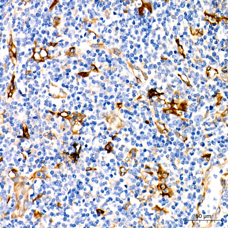
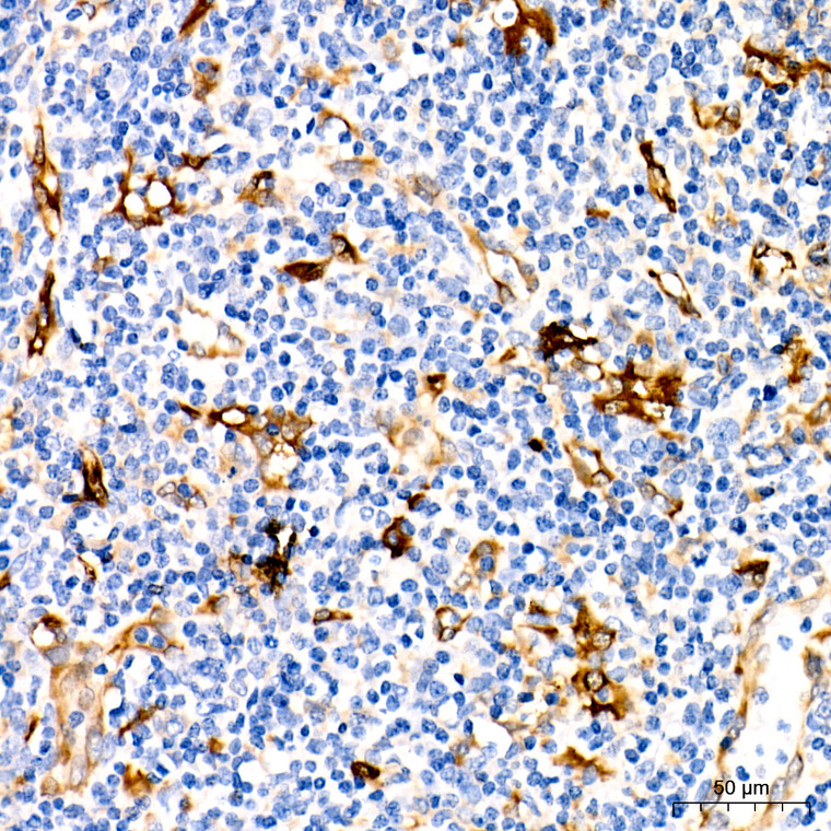
| Host: |
Rabbit |
| Applications: |
WB/IHC |
| Reactivity: |
Human/Mouse/Rat |
| Note: |
STRICTLY FOR FURTHER SCIENTIFIC RESEARCH USE ONLY (RUO). MUST NOT TO BE USED IN DIAGNOSTIC OR THERAPEUTIC APPLICATIONS. |
| Short Description: |
Rabbit monoclonal antibody anti-Fascin (249-362) is suitable for use in Western Blot and Immunohistochemistry research applications. |
| Clonality: |
Monoclonal |
| Clone ID: |
S5MR |
| Conjugation: |
Unconjugated |
| Isotype: |
IgG |
| Formulation: |
PBS with 0.02% Sodium Azide, 0.05% BSA, 50% Glycerol, pH7.3. |
| Purification: |
Affinity purification |
| Dilution Range: |
WB 1:500-1:1000IHC-P 1:50-1:200 |
| Storage Instruction: |
Store at-20°C for up to 1 year from the date of receipt, and avoid repeat freeze-thaw cycles. |
| Gene Symbol: |
FSCN1 |
| Gene ID: |
6624 |
| Uniprot ID: |
FSCN1_HUMAN |
| Immunogen Region: |
249-362 |
| Immunogen: |
Recombinant fusion protein containing a sequence corresponding to amino acids 249-362 of human Fascin/FSCN1 (Q16658). |
| Immunogen Sequence: |
GKDELFALEQSCAQVVLQAA NERNVSTRQGMDLSANQDEE TDQETFQLEIDRDTKKCAFR THTGKYWTLTATGGVQSTAS SKNASCYFDIEWRDRRITLR ASNGKFVTSKKNGQ |
| Tissue Specificity | Ubiquitous. |
| Post Translational Modifications | Phosphorylation at Ser-39 inhibits actin-binding. Phosphorylation is required for the reorganization of the actin cytoskeleton in response to NGF. |
| Function | Actin-binding protein that contains 2 major actin binding sites. Organizes filamentous actin into parallel bundles. Plays a role in the organization of actin filament bundles and the formation of microspikes, membrane ruffles, and stress fibers. Important for the formation of a diverse set of cell protrusions, such as filopodia, and for cell motility and migration. Mediates reorganization of the actin cytoskeleton and axon growth cone collapse in response to NGF. |
| Protein Name | Fascin55 Kda Actin-Bundling ProteinSinged-Like ProteinP55 |
| Database Links | Reactome: R-HSA-6785807 |
| Cellular Localisation | CytoplasmCytosolCell CortexCytoskeletonStress FiberCell ProjectionFilopodiumInvadopodiumMicrovillusCell JunctionColocalized With Rufy3 And F-Actin At Filipodia Of The Axonal Growth ConeColocalized With Dbn1 And F-Actin At The Transitional Domain Of The Axonal Growth Cone |
| Alternative Antibody Names | Anti-Fascin antibodyAnti-55 Kda Actin-Bundling Protein antibodyAnti-Singed-Like Protein antibodyAnti-P55 antibodyAnti-FSCN1 antibodyAnti-FAN1 antibodyAnti-HSN antibodyAnti-SNL antibody |
Information sourced from Uniprot.org
12 months for antibodies. 6 months for ELISA Kits. Please see website T&Cs for further guidance


