| Host: |
Rabbit |
| Applications: |
WB/IHC/IF |
| Reactivity: |
Human/Mouse/Rat |
| Note: |
STRICTLY FOR FURTHER SCIENTIFIC RESEARCH USE ONLY (RUO). MUST NOT TO BE USED IN DIAGNOSTIC OR THERAPEUTIC APPLICATIONS. |
| Short Description: |
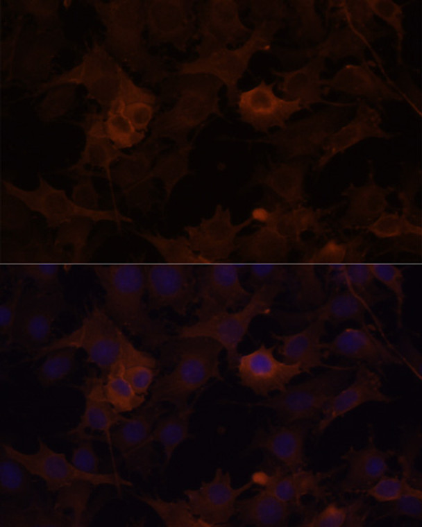
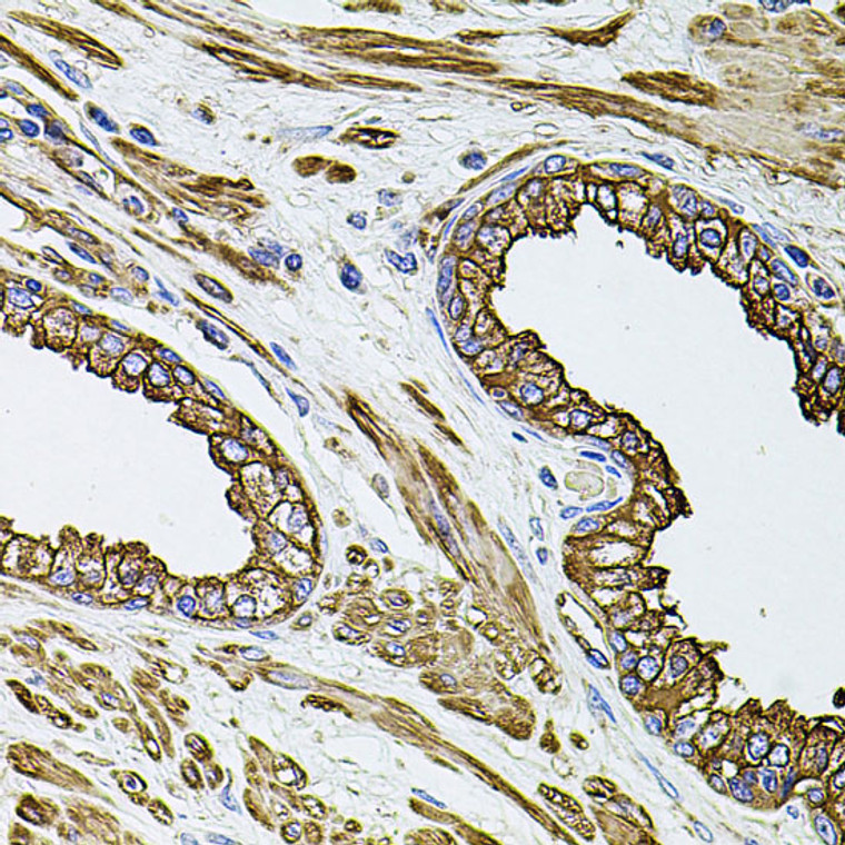
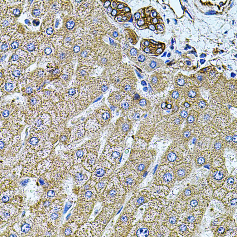
Rabbit polyclonal antibody anti-FLNB (1686-1785) is suitable for use in Western Blot, Immunohistochemistry and Immunofluorescence research applications. |
| Clonality: |
Polyclonal |
| Conjugation: |
Unconjugated |
| Isotype: |
IgG |
| Formulation: |
PBS with 0.02% Sodium Azide, 50% Glycerol, pH7.3. |
| Purification: |
Affinity purification |
| Dilution Range: |
WB 1:500-1:1000IHC-P 1:50-1:100IF/ICC 1:20-1:100 |
| Storage Instruction: |
Store at-20°C for up to 1 year from the date of receipt, and avoid repeat freeze-thaw cycles. |
| Gene Symbol: |
FLNB |
| Gene ID: |
2317 |
| Uniprot ID: |
FLNB_HUMAN |
| Immunogen Region: |
1686-1785 |
| Immunogen: |
Recombinant fusion protein containing a sequence corresponding to amino acids 1686-1785 of human FLNB (NP_001157789.1). |
| Immunogen Sequence: |
PDGTEAEADVIENEDGTYDI FYTAAKPGTYVIYVRFGGVD IPNSPFTVMATDGEVTAVEE APVNACPPGFRPWVTEEAYV PVSDMNGLGFKPFDLVIPFA |
| Tissue Specificity | Ubiquitous. Isoform 1 and isoform 2 are expressed in placenta, bone marrow, brain, umbilical vein endothelial cells (HUVEC), retina and skeletal muscle. Isoform 1 is predominantly expressed in prostate, uterus, liver, thyroid, stomach, lymph node, small intestine, spleen, skeletal muscle, kidney, placenta, pancreas, heart, lung, platelets, endothelial cells, megakaryocytic and erythroleukemic cell lines. Isoform 2 is predominantly expressed in spinal cord, platelet and Daudi cells. Also expressed in thyroid adenoma, neurofibrillary tangles (NFT), senile plaques in the hippocampus and cerebral cortex in Alzheimer disease (AD). Isoform 3 and isoform 6 are expressed predominantly in lung, heart, skeletal muscle, testis, spleen, thymus and leukocytes. Isoform 4 and isoform 5 are expressed in heart. |
| Post Translational Modifications | ISGylation prevents ability to interact with the upstream activators of the JNK cascade and inhibits IFNA-induced JNK signaling. Ubiquitination by a SCF-like complex containing ASB2 isoform 1 leads to proteasomal degradation which promotes muscle differentiation. |
| Function | Connects cell membrane constituents to the actin cytoskeleton. May promote orthogonal branching of actin filaments and links actin filaments to membrane glycoproteins. Anchors various transmembrane proteins to the actin cytoskeleton. Interaction with FLNA may allow neuroblast migration from the ventricular zone into the cortical plate. Various interactions and localizations of isoforms affect myotube morphology and myogenesis. Isoform 6 accelerates muscle differentiation in vitro. |
| Protein Name | Filamin-BFln-BAbp-278Abp-280 HomologActin-Binding-Like ProteinBeta-FilaminFilamin Homolog 1Fh1Filamin-3Thyroid AutoantigenTruncated Actin-Binding ProteinTruncated Abp |
| Database Links | Reactome: R-HSA-1169408 |
| Cellular Localisation | Isoform 1: CytoplasmCell CortexCytoplasmCytoskeletonStress FiberMyofibrilSarcomereZ LineIn Differentiating MyotubesIsoform 1Isoform 2 And Isoform 3 Are Localized Diffusely Throughout The Cytoplasm With Regions Of Enrichment At The Longitudinal Actin Stress FiberIn Differentiated TubesIsoform 1 Is Also Detected Within The Z-LinesIsoform 2: CytoplasmIsoform 3: CytoplasmIsoform 6: CytoplasmPolarized At The Periphery Of Myotubes |
| Alternative Antibody Names | Anti-Filamin-B antibodyAnti-Fln-B antibodyAnti-Abp-278 antibodyAnti-Abp-280 Homolog antibodyAnti-Actin-Binding-Like Protein antibodyAnti-Beta-Filamin antibodyAnti-Filamin Homolog 1 antibodyAnti-Fh1 antibodyAnti-Filamin-3 antibodyAnti-Thyroid Autoantigen antibodyAnti-Truncated Actin-Binding Protein antibodyAnti-Truncated Abp antibodyAnti-FLNB antibodyAnti-FLN1L antibodyAnti-FLN3 antibodyAnti-TABP antibodyAnti-TAP antibody |
Information sourced from Uniprot.org
12 months for antibodies. 6 months for ELISA Kits. Please see website T&Cs for further guidance


