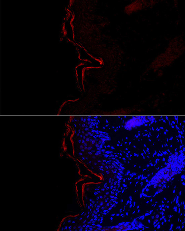
| Host: |
Rabbit |
| Applications: |
WB/IF |
| Reactivity: |
Human/Mouse/Rat |
| Note: |
STRICTLY FOR FURTHER SCIENTIFIC RESEARCH USE ONLY (RUO). MUST NOT TO BE USED IN DIAGNOSTIC OR THERAPEUTIC APPLICATIONS. |
| Short Description: |
Rabbit polyclonal antibody anti-FLG (1-92) is suitable for use in Western Blot and Immunofluorescence research applications. |
| Clonality: |
Polyclonal |
| Conjugation: |
Unconjugated |
| Isotype: |
IgG |
| Formulation: |
PBS with 0.01% Thimerosal, 50% Glycerol, pH7.3. |
| Purification: |
Affinity purification |
| Dilution Range: |
WB 1:1000-1:5000IF/ICC 1:50-1:100 |
| Storage Instruction: |
Store at-20°C for up to 1 year from the date of receipt, and avoid repeat freeze-thaw cycles. |
| Gene Symbol: |
FLG |
| Gene ID: |
2312 |
| Uniprot ID: |
FILA_HUMAN |
| Immunogen Region: |
1-92 |
| Immunogen: |
Recombinant fusion protein containing a sequence corresponding to amino acids 1-92 of human FLG (NP_002007.1). |
| Immunogen Sequence: |
MSTLLENIFAIINLFKQYSK KDKNTDTLSKKELKELLEKE FRQILKNPDDPDMVDVFMDH LDIDHNKKIDFTEFLLMVFK LAQAYYESTRKE |
| Tissue Specificity | Expressed in skin, thymus, stomach, tonsils, testis, placenta, kidney, pancreas, mammary gland, bladder, thyroid, salivary gland and trachea, but not detected in heart, brain, liver, lung, bone marrow, small intestine, spleen, prostate, colon, or adrenal gland. In the skin, mainly expressed in stratum granulosum of the epidermis. |
| Post Translational Modifications | Filaggrin is initially synthesized as a large, insoluble, highly phosphorylated precursor containing many tandem copies of 324 AA, which are not separated by large linker sequences. During terminal differentiation it is dephosphorylated and proteolytically cleaved. The N-terminal of the mature protein is heterogeneous, and is blocked by the formation of pyroglutamate. Undergoes deimination of some arginine residues (citrullination). |
| Function | Aggregates keratin intermediate filaments and promotes disulfide-bond formation among the intermediate filaments during terminal differentiation of mammalian epidermis. |
| Protein Name | Filaggrin |
| Database Links | Reactome: R-HSA-6809371 |
| Cellular Localisation | Cytoplasmic GranuleIn The Stratum Granulosum Of The EpidermisLocalized Within Keratohyalin GranulesIn Granular Keratinocytes And In Lower CorneocytesColocalizes With Calpain-1/Capn1 |
| Alternative Antibody Names | Anti-Filaggrin antibodyAnti-FLG antibody |
Information sourced from Uniprot.org
12 months for antibodies. 6 months for ELISA Kits. Please see website T&Cs for further guidance


