| Host: |
Rabbit |
| Applications: |
WB |
| Reactivity: |
Human |
| Note: |
STRICTLY FOR FURTHER SCIENTIFIC RESEARCH USE ONLY (RUO). MUST NOT TO BE USED IN DIAGNOSTIC OR THERAPEUTIC APPLICATIONS. |
| Short Description: |
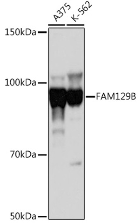
Rabbit polyclonal antibody anti-FAM129B (447-746) is suitable for use in Western Blot research applications. |
| Clonality: |
Polyclonal |
| Conjugation: |
Unconjugated |
| Isotype: |
IgG |
| Formulation: |
PBS with 0.01% Thimerosal, 50% Glycerol, pH7.3. |
| Purification: |
Affinity purification |
| Dilution Range: |
WB 1:500-1:1000 |
| Storage Instruction: |
Store at-20°C for up to 1 year from the date of receipt, and avoid repeat freeze-thaw cycles. |
| Gene Symbol: |
NIBAN2 |
| Gene ID: |
64855 |
| Uniprot ID: |
NIBA2_HUMAN |
| Immunogen Region: |
447-746 |
| Immunogen: |
Recombinant fusion protein containing a sequence corresponding to amino acids 447-746 of human FAM129B (NP_073744.2). |
| Immunogen Sequence: |
TFETLLHQELGKGPTKEELC KSIQRVLERVLKKYDYDSSS VRKRFFREALLQISIPFLLK KLAPTCKSELPRFQELIFED FARFILVENTYEEVVLQTVM KDILQAVKEAAVQRKHNLYR DSMVMHNSDPNLHLLAEGAP IDWGEEYSNSGGGGSPSPST PESATLSEKRRRAKQVVSVV QDEEVGLPFEASPESPPPAS PDGVTEIRGLLAQGLRPESP PPAGPLLNGAPAGESPQPK |
| Post Translational Modifications | Phosphorylated at Ser-641, Ser-646, Ser-692 and Ser-696 by the BRAF/MKK/ERK signaling cascade. In melanoma cells, the C-terminal phosphorylation may prevent targeting to the plasma membrane. As apoptosis proceeds, degraded via an proteasome-independent pathway, probably by caspases. |
| Function | May play a role in apoptosis suppression. May promote melanoma cell invasion in vitro. |
| Protein Name | Protein Niban 2Meg-3Melanoma Invasion By ErkMinervaNiban-Like Protein 1Protein Fam129b |
| Cellular Localisation | CytoplasmCytosolCell JunctionAdherens JunctionMembraneLipid-AnchorIn Exponentially Growing CellsExclusively CytoplasmicCell Membrane Localization Is Observed When Cells Reach Confluency And During TelophaseIn Melanoma CellsTargeting To The Plasma Membrane May Be Impaired By C-Terminal Phosphorylation |
| Alternative Antibody Names | Anti-Protein Niban 2 antibodyAnti-Meg-3 antibodyAnti-Melanoma Invasion By Erk antibodyAnti-Minerva antibodyAnti-Niban-Like Protein 1 antibodyAnti-Protein Fam129b antibodyAnti-NIBAN2 antibodyAnti-C9orf88 antibodyAnti-FAM129B antibody |
Information sourced from Uniprot.org
12 months for antibodies. 6 months for ELISA Kits. Please see website T&Cs for further guidance


