| Host: |
Rabbit |
| Applications: |
WB |
| Reactivity: |
Human/Mouse/Rat |
| Note: |
STRICTLY FOR FURTHER SCIENTIFIC RESEARCH USE ONLY (RUO). MUST NOT TO BE USED IN DIAGNOSTIC OR THERAPEUTIC APPLICATIONS. |
| Short Description: |
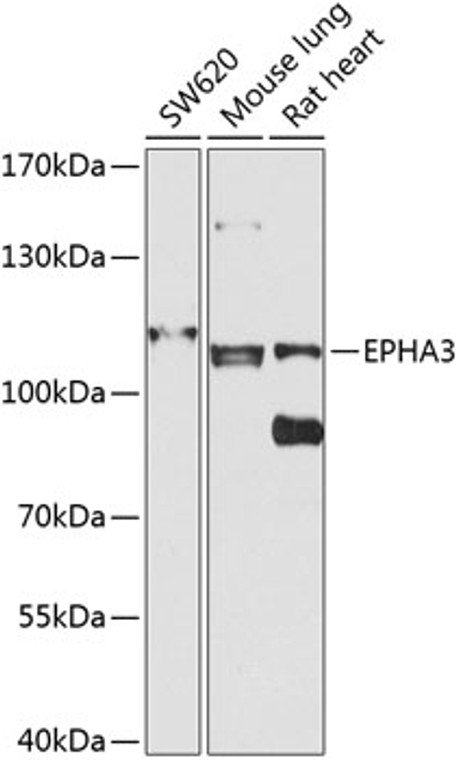
Rabbit polyclonal antibody anti-EPHA3 (400-545) is suitable for use in Western Blot research applications. |
| Clonality: |
Polyclonal |
| Conjugation: |
Unconjugated |
| Isotype: |
IgG |
| Formulation: |
PBS with 0.02% Sodium Azide, 50% Glycerol, pH7.3. |
| Purification: |
Affinity purification |
| Dilution Range: |
WB 1:500-1:2000 |
| Storage Instruction: |
Store at-20°C for up to 1 year from the date of receipt, and avoid repeat freeze-thaw cycles. |
| Gene Symbol: |
EPHA3 |
| Gene ID: |
2042 |
| Uniprot ID: |
EPHA3_HUMAN |
| Immunogen Region: |
400-545 |
| Immunogen: |
Recombinant fusion protein containing a sequence corresponding to amino acids 400-545 of human EPHA3 (NP_005224.2). |
| Immunogen Sequence: |
LAHTNYTFEIDAVNGVSELS SPPRQFAAVSITTNQAAPSP VLTIKKDRTSRNSISLSWQE PEHPNGIILDYEVKYYEKQE QETSYTILRARGTNVTISSL KPDTIYVFQIRARTAAGYGT NSRKFEFETSPDSFSISGES SQVVMI |
| Tissue Specificity | Widely expressed. Highest level in placenta. |
| Post Translational Modifications | Autophosphorylates upon activation by EFNA5. Phosphorylation on Tyr-602 mediates interaction with NCK1. Dephosphorylated by PTPN1. |
| Function | Receptor tyrosine kinase which binds promiscuously membrane-bound ephrin family ligands residing on adjacent cells, leading to contact-dependent bidirectional signaling into neighboring cells. The signaling pathway downstream of the receptor is referred to as forward signaling while the signaling pathway downstream of the ephrin ligand is referred to as reverse signaling. Highly promiscuous for ephrin-A ligands it binds preferentially EFNA5. Upon activation by EFNA5 regulates cell-cell adhesion, cytoskeletal organization and cell migration. Plays a role in cardiac cells migration and differentiation and regulates the formation of the atrioventricular canal and septum during development probably through activation by EFNA1. Involved in the retinotectal mapping of neurons. May also control the segregation but not the guidance of motor and sensory axons during neuromuscular circuit development. |
| Protein Name | Ephrin Type-A Receptor 3Eph-Like Kinase 4Ek4Hek4HekHuman Embryo KinaseTyrosine-Protein Kinase Tyro4Tyrosine-Protein Kinase Receptor Etk1Eph-Like Tyrosine Kinase 1 |
| Database Links | Reactome: R-HSA-2682334Reactome: R-HSA-3928663Reactome: R-HSA-3928665 |
| Cellular Localisation | Isoform 1: Cell MembraneSingle-Pass Type I Membrane ProteinIsoform 2: Secreted |
| Alternative Antibody Names | Anti-Ephrin Type-A Receptor 3 antibodyAnti-Eph-Like Kinase 4 antibodyAnti-Ek4 antibodyAnti-Hek4 antibodyAnti-Hek antibodyAnti-Human Embryo Kinase antibodyAnti-Tyrosine-Protein Kinase Tyro4 antibodyAnti-Tyrosine-Protein Kinase Receptor Etk1 antibodyAnti-Eph-Like Tyrosine Kinase 1 antibodyAnti-EPHA3 antibodyAnti-ETK antibodyAnti-ETK1 antibodyAnti-HEK antibodyAnti-TYRO4 antibody |
Information sourced from Uniprot.org
12 months for antibodies. 6 months for ELISA Kits. Please see website T&Cs for further guidance


