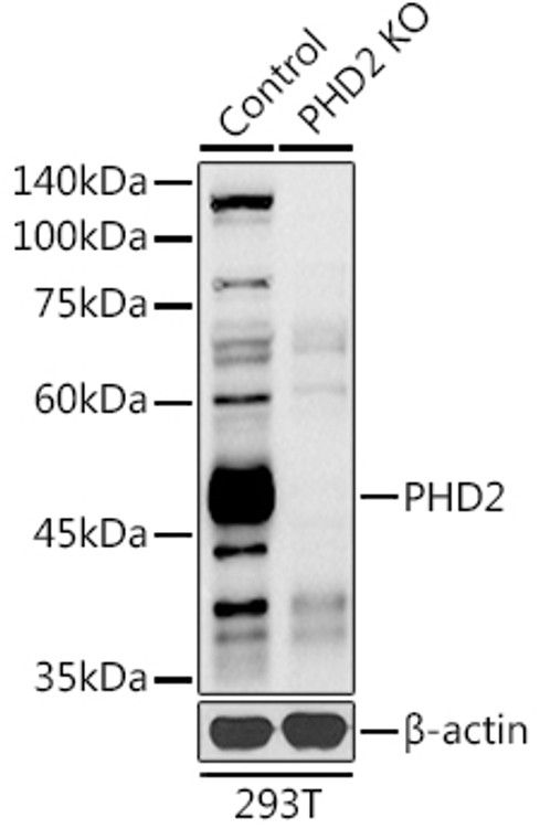
| Host: |
Rabbit |
| Applications: |
WB/IHC/IF |
| Reactivity: |
Human/Mouse/Rat |
| Note: |
STRICTLY FOR FURTHER SCIENTIFIC RESEARCH USE ONLY (RUO). MUST NOT TO BE USED IN DIAGNOSTIC OR THERAPEUTIC APPLICATIONS. |
| Short Description: |
Rabbit polyclonal antibody anti-PHD2 (1-100) is suitable for use in Western Blot, Immunohistochemistry and Immunofluorescence research applications. |
| Clonality: |
Polyclonal |
| Conjugation: |
Unconjugated |
| Isotype: |
IgG |
| Formulation: |
PBS with 0.05% Proclin300, 50% Glycerol, pH7.3. |
| Purification: |
Affinity purification |
| Dilution Range: |
WB 1:500-1:1000IHC-P 1:50-1:200IF/ICC 1:50-1:200 |
| Storage Instruction: |
Store at-20°C for up to 1 year from the date of receipt, and avoid repeat freeze-thaw cycles. |
| Gene Symbol: |
EGLN1 |
| Gene ID: |
54583 |
| Uniprot ID: |
EGLN1_HUMAN |
| Immunogen Region: |
1-100 |
| Immunogen: |
A synthetic peptide corresponding to a sequence within amino acids 1-100 of human PHD2/EGLN1 (NP_071334.1). |
| Immunogen Sequence: |
MANDSGGPGGPSPSERDRQY CELCGKMENLLRCSRCRSSF YCCKEHQRQDWKKHKLVCQG SEGALGHGVGPHQHSGPAPP AAVPPPRAGAREPRKAAARR |
| Tissue Specificity | widely expressed with highest levels in skeletal muscle and heart, moderate levels in pancreas, brain (dopaminergic neurons of adult and fetal substantia nigra) and kidney, and lower levels in lung and liver. widely expressed with highest levels in brain, kidney and adrenal gland. Expressed in cardiac myocytes, aortic endothelial cells and coronary artery smooth muscle. .expressed in adult and fetal heart, brain, liver, lung, skeletal muscle and kidney. Also expressed in placenta. Highest levels in adult heart, brain, lung and liver and fetal brain, heart spleen and skeletal muscle. |
| Post Translational Modifications | S-nitrosylation inhibits the enzyme activity up to 60% under aerobic conditions. Chelation of Fe(2+) has no effect on the S-nitrosylation. It is uncertain whether nitrosylation occurs on Cys-323 or Cys-326. |
| Function | Cellular oxygen sensor that catalyzes, under normoxic conditions, the post-translational formation of 4-hydroxyproline in hypoxia-inducible factor (HIF) alpha proteins. Hydroxylates a specific proline found in each of the oxygen-dependent degradation (ODD) domains (N-terminal, NODD, and C-terminal, CODD) of HIF1A. Also hydroxylates HIF2A. Has a preference for the CODD site for both HIF1A and HIF1B. Hydroxylated HIFs are then targeted for proteasomal degradation via the von Hippel-Lindau ubiquitination complex. Under hypoxic conditions, the hydroxylation reaction is attenuated allowing HIFs to escape degradation resulting in their translocation to the nucleus, heterodimerization with HIF1B, and increased expression of hypoxy-inducible genes. EGLN1 is the most important isozyme under normoxia and, through regulating the stability of HIF1, involved in various hypoxia-influenced processes such as angiogenesis in retinal and cardiac functionality. Target proteins are preferentially recognized via a LXXLAP motif. |
| Protein Name | Egl Nine Homolog 1Hypoxia-Inducible Factor Prolyl Hydroxylase 2Hif-Ph2Hif-Prolyl Hydroxylase 2Hph-2Prolyl Hydroxylase Domain-Containing Protein 2Phd2Sm-20 |
| Database Links | Reactome: R-HSA-1234176 |
| Cellular Localisation | CytoplasmNucleusMainly CytoplasmicShuttles Between The Nucleus And CytoplasmNuclear Export Requires Functional Xpo1 |
| Alternative Antibody Names | Anti-Egl Nine Homolog 1 antibodyAnti-Hypoxia-Inducible Factor Prolyl Hydroxylase 2 antibodyAnti-Hif-Ph2 antibodyAnti-Hif-Prolyl Hydroxylase 2 antibodyAnti-Hph-2 antibodyAnti-Prolyl Hydroxylase Domain-Containing Protein 2 antibodyAnti-Phd2 antibodyAnti-Sm-20 antibodyAnti-EGLN1 antibodyAnti-C1orf12 antibodyAnti-PNAS-118 antibodyAnti-PNAS-137 antibody |
Information sourced from Uniprot.org
12 months for antibodies. 6 months for ELISA Kits. Please see website T&Cs for further guidance

![Immunofluorescence analysis of NIH/3T3 cells using [KO Validated] PHD2/EGLN1 Rabbit polyclonal antibody (STJ116768) at dilution of 1:100 (40x lens). Blue: DAPI for nuclear staining.](https://cdn11.bigcommerce.com/s-zso2xnchw9/images/stencil/760x760/products/95742/377887/STJ116768_1__81838.1713145295.jpg?c=1)
![Immunohistochemistry analysis of paraffin-embedded rat heart using [KO Validated] PHD2/EGLN1 Rabbit polyclonal antibody (STJ116768) at dilution of 1:100 (40x lens). Perform high pressure antigen retrieval with 10 mM citrate buffer pH 6. 0 before commencing with immunohistochemistry staining protocol.](https://cdn11.bigcommerce.com/s-zso2xnchw9/images/stencil/760x760/products/95742/377888/STJ116768_2__25558.1713145295.jpg?c=1)
![Immunohistochemistry analysis of paraffin-embedded rat brain using [KO Validated] PHD2/EGLN1 Rabbit polyclonal antibody (STJ116768) at dilution of 1:100 (40x lens). Perform high pressure antigen retrieval with 10 mM citrate buffer pH 6. 0 before commencing with immunohistochemistry staining protocol.](https://cdn11.bigcommerce.com/s-zso2xnchw9/images/stencil/760x760/products/95742/377889/STJ116768_3__71580.1713145296.jpg?c=1)
![Immunohistochemistry analysis of paraffin-embedded mouse brain using [KO Validated] PHD2/EGLN1 Rabbit polyclonal antibody (STJ116768) at dilution of 1:100 (40x lens). Perform high pressure antigen retrieval with 10 mM citrate buffer pH 6. 0 before commencing with immunohistochemistry staining protocol.](https://cdn11.bigcommerce.com/s-zso2xnchw9/images/stencil/760x760/products/95742/377890/STJ116768_4__40393.1713145298.jpg?c=1)


