| Host: |
Rabbit |
| Applications: |
WB/IF |
| Reactivity: |
Human |
| Note: |
STRICTLY FOR FURTHER SCIENTIFIC RESEARCH USE ONLY (RUO). MUST NOT TO BE USED IN DIAGNOSTIC OR THERAPEUTIC APPLICATIONS. |
| Short Description: |
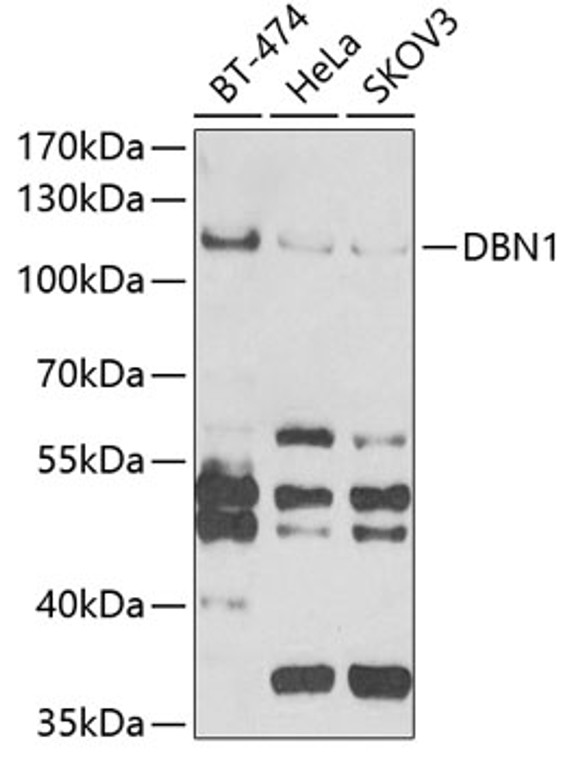
Rabbit polyclonal antibody anti-DBN1 (350-649) is suitable for use in Western Blot and Immunofluorescence research applications. |
| Clonality: |
Polyclonal |
| Conjugation: |
Unconjugated |
| Isotype: |
IgG |
| Formulation: |
PBS with 0.02% Sodium Azide, 50% Glycerol, pH7.3. |
| Purification: |
Affinity purification |
| Dilution Range: |
WB 1:500-1:2000IF/ICC 1:50-1:100 |
| Storage Instruction: |
Store at-20°C for up to 1 year from the date of receipt, and avoid repeat freeze-thaw cycles. |
| Gene Symbol: |
DBN1 |
| Gene ID: |
1627 |
| Uniprot ID: |
DREB_HUMAN |
| Immunogen Region: |
350-649 |
| Immunogen: |
Recombinant fusion protein containing a sequence corresponding to amino acids 350-649 of human DBN1 (NP_004386.2). |
| Immunogen Sequence: |
EQIERALDEVTSSQPPPLPP PPPPAQETQEPSPILDSEET RAAAPQAWAGPMEEPPQAQA PPRGPGSPAEDLMFMESAEQ AVLAAPVEPATADATEIHDA ADTIETDTATADTTVANNVP PAATSLIDLWPGNGEGASTL QGEPRAPTPPSGTEVTLAEV PLLDEVAPEPLLPAGEGCAT LLNFDELPEPPATFCDPEEV EGEPLAAPQTPTLPSALEEL EQEQEPEPHLLTNGETTQK |
| Tissue Specificity | Expressed in the brain, with expression in the molecular layer of the dentate gyrus, stratum pyramidale, and stratum radiatum of the hippocampus (at protein level). Also expressed in the terminal varicosities distributed along dendritic trees of pyramidal cells in CA4 and CA3 of the hippocampus (at protein level). Expressed in pyramidal cells in CA2, CA1 and the subiculum of the hippocampus (at protein level). Expressed in peripheral blood lymphocytes, including T-cells (at protein level). Expressed in the brain. Expressed in the heart, placenta, lung, skeletal muscle, kidney, pancreas, skin fibroblasts, gingival fibroblasts and bone-derived cells (Ref.2). |
| Function | Actin cytoskeleton-organizing protein that plays a role in the formation of cell projections. Required for actin polymerization at immunological synapses (IS) and for the recruitment of the chemokine receptor CXCR4 to IS. Plays a role in dendritic spine morphogenesis and organization, including the localization of the dopamine receptor DRD1 to the dendritic spines. Involved in memory-related synaptic plasticity in the hippocampus. |
| Protein Name | DrebrinDevelopmentally-Regulated Brain Protein |
| Database Links | Reactome: R-HSA-9013405Reactome: R-HSA-9013407Reactome: R-HSA-9013418Reactome: R-HSA-9013422 |
| Cellular Localisation | CytoplasmCell ProjectionDendriteCell CortexCell JunctionGrowth ConeIn The Absence Of AntigenEvenly Distributed Throughout Subcortical Regions Of The T-Cell Membrane And CytoplasmIn The Presence Of AntigenDistributes To The Immunological Synapse Forming At The T-Cell-Apc Contact AreaWhere It Localizes At The Peripheral And Distal Supramolecular Activation Clusters (Smac)Colocalized With Rufy3 And F-Actin At The Transitional Domain Of The Axonal Growth Cone |
| Alternative Antibody Names | Anti-Drebrin antibodyAnti-Developmentally-Regulated Brain Protein antibodyAnti-DBN1 antibodyAnti-D0S117E antibody |
Information sourced from Uniprot.org
12 months for antibodies. 6 months for ELISA Kits. Please see website T&Cs for further guidance


