• Western blot analysis of lysate from HeLa cells, using IL8 Antibody.
  • Immunohistochemical analysis of paraffin-embedded human-skin, antibody was diluted at 1:100
  • Immunohistochemical analysis of paraffin-embedded human-skin, antibody was diluted at 1:100
  • Immunohistochemical analysis of paraffin-embedded Human-kidney tissue. 1, IL-8 Polyclonal Antibody was diluted at 1:200 (4°C, overnight). 2, Sodium citrate pH 6.0 was used for antibody retrieval (>98°C, 20min). 3, Secondary antibody was diluted at 1:200 (room tempeRature, 30min). Negative control was used by secondary antibody only.
  • Western blot analysis of HeLa cells using IL-8 Polyclonal Antibody.. Secondary antibody was diluted at 1:20000
  • Western blot analysis of various cells using primary antibody diluted at 1:1000 (4°C overnight). Secondary antibody:Goat Anti-rabbit IgG IRDye 800 ( diluted at 1:5000, 25°C, 1 hour). Cell lysate was extracted by Minute Plasma Membrane Protein Isolation and Cell Fractionation Kit (SM-005, Inventbiotech, MN, USA).
  • Immunohistochemical analysis of paraffin-embedded Human-liver-cancer tissue. 1, IL-8 Polyclonal Antibody was diluted at 1:200 (4°C, overnight). 2, Sodium citrate pH 6.0 was used for antibody retrieval (>98°C, 20min). 3, Secondary antibody was diluted at 1:200 (room tempeRature, 30min). Negative control was used by secondary antibody only.
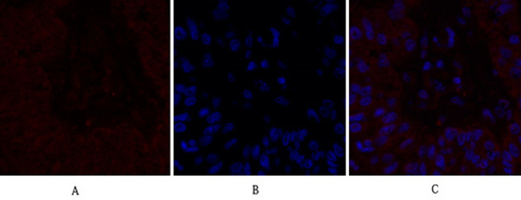
  • Immunofluorescence analysis of human-kidney tissue. 1, IL-8 Polyclonal Antibody (red) was diluted at 1:200 (4°C, overnight). 2, Cy3 labled Secondary antibody was diluted at 1:300 (room temperature, 50min).3, Picture B: DAPI (blue) 10min. Picture A:Target. Picture B: DAPI. Picture C: merge of A+B
  • Immunofluorescence analysis of human-kidney tissue. 1, IL-8 Polyclonal Antibody (red) was diluted at 1:200 (4°C, overnight). 2, Cy3 labled Secondary antibody was diluted at 1:300 (room temperature, 50min).3, Picture B: DAPI (blue) 10min. Picture A:Target. Picture B: DAPI. Picture C: merge of A+B
  • Immunofluorescence analysis of human-liver-cancer tissue. 1, IL-8 Polyclonal Antibody (red) was diluted at 1:200 (4°C, overnight). 2, Cy3 labled Secondary antibody was diluted at 1:300 (room temperature, 50min).3, Picture B: DAPI (blue) 10min. Picture A:Target. Picture B: DAPI. Picture C: merge of A+B
  • Immunofluorescence analysis of human-liver-cancer tissue. 1, IL-8 Polyclonal Antibody (red) was diluted at 1:200 (4°C, overnight). 2, Cy3 labled Secondary antibody was diluted at 1:300 (room temperature, 50min).3, Picture B: DAPI (blue) 10min. Picture A:Target. Picture B: DAPI. Picture C: merge of A+B
  • Immunofluorescence analysis of human-breast-cancer tissue. 1, IL-8 Polyclonal Antibody (red) was diluted at 1:200 (4°C, overnight). 2, Cy3 labled Secondary antibody was diluted at 1:300 (room temperature, 50min).3, Picture B: DAPI (blue) 10min. Picture A:Target. Picture B: DAPI. Picture C: merge of A+B
  • Immunofluorescence analysis of human-breast-cancer tissue. 1, IL-8 Polyclonal Antibody (red) was diluted at 1:200 (4°C, overnight). 2, Cy3 labled Secondary antibody was diluted at 1:300 (room temperature, 50min).3, Picture B: DAPI (blue) 10min. Picture A:Target. Picture B: DAPI. Picture C: merge of A+B
  • Immunofluorescence analysis of Hela cell. 1, IL-8 Polyclonal Antibody (green) was diluted at 1:200 (4°C overnight). 2, Goat Anti Rabbit Alexa Fluor 488 Catalog: (NA was diluted at 1:1000 (room temperature, 50min). 3 DAPI (blue) 10min.

Anti-CXCL8 antibody (50-99 aa) (STJ96518)

SKU:
STJ96518

Current Stock:
Host: Rabbit
Applications: IF/WB/IHC/ELISA
Reactivity: Human
Note: STRICTLY FOR FURTHER SCIENTIFIC RESEARCH USE ONLY (RUO). MUST NOT TO BE USED IN DIAGNOSTIC OR THERAPEUTIC APPLICATIONS.
Short Description: Rabbit polyclonal antibody anti-Interleukin-8 ligand 8 (50-99 aa) is suitable for use in Immunofluorescence, Western Blot, Immunohistochemistry and ELISA research applications.
Clonality: Polyclonal
Conjugation: Unconjugated
Isotype: IgG
Formulation: Liquid in PBS containing 50% Glycerol, 0.5% BSA and 0.02% Sodium Azide.
Purification: The antibody was affinity-purified from rabbit antiserum by affinity-chromatography using epitope-specific immunogen.
Concentration: 1 mg/mL
Dilution Range: IF 1:50-200
WB 1:500-1:2000
IHC-P 1:100-300
ELISA 1:20000
Storage Instruction: Store at-20°C for up to 1 year from the date of receipt, and avoid repeat freeze-thaw cycles.
Gene Symbol: CXCL8
Gene ID: 3576
Uniprot ID: IL8_HUMAN
Immunogen Region: 50-99 aa
Specificity: IL-8 Polyclonal Antibody detects endogenous levels of IL-8 protein.
Immunogen: The antiserum was produced against synthesized peptide derived from the C-terminal region of human IL8 at the amino acid range 50-99
Function Chemotactic factor that mediates inflammatory response by attracting neutrophils, basophils, and T-cells to clear pathogens and protect the host from infection. Also plays an important role in neutrophil activation. Released in response to an inflammatory stimulus, exerts its effect by binding to the G-protein-coupled receptors CXCR1 and CXCR2, primarily found in neutrophils, monocytes and endothelial cells. G-protein heterotrimer (alpha, beta, gamma subunits) constitutively binds to CXCR1/CXCR2 receptor and activation by IL8 leads to beta and gamma subunits release from Galpha (GNAI2 in neutrophils) and activation of several downstream signaling pathways including PI3K and MAPK pathways.
Protein Name Interleukin-8
Il-8
C-X-C Motif Chemokine 8
Chemokine
C-X-C Motif Ligand 8
Emoctakin
Granulocyte Chemotactic Protein 1
Gcp-1
Monocyte-Derived Neutrophil Chemotactic Factor
Mdncf
Monocyte-Derived Neutrophil-Activating Peptide
Monap
Neutrophil-Activating Protein 1
Nap-1
Protein 3-10c
T-Cell Chemotactic Factor Cleaved Into - Mdncf-A
Gcp/Il-8 Protein Iv
Il8/Nap1 Form I - Interleukin-8
(Ala-Il-877
Gcp/Il-8 Protein Ii
Il-8(1-77
Il8/Nap1 Form Ii
Mdncf-B - Il-8(5-77 - Il-8(6-77
(Ser-Il-872
Gcp/Il-8 Protein I
Il8/Nap1 Form Iii
Lymphocyte-Derived Neutrophil-Activating Factor
Lynap
Mdncf-C
Neutrophil-Activating Factor
Naf - Il-8(7-77
Gcp/Il-8 Protein V
Il8/Nap1 Form Iv - Il-8(8-77
Gcp/Il-8 Protein Vi
Il8/Nap1 Form V - Il-8(9-77
Gcp/Il-8 Protein Iii
Il8/Nap1 Form Vi
Database Links Reactome: R-HSA-2559582
Reactome: R-HSA-375276
Reactome: R-HSA-380108
Reactome: R-HSA-380994
Reactome: R-HSA-418594
Reactome: R-HSA-6783783
Reactome: R-HSA-6785807
Cellular Localisation Secreted
Alternative Antibody Names Anti-Interleukin-8 antibody
Anti-Il-8 antibody
Anti-C-X-C Motif Chemokine 8 antibody
Anti-Chemokine antibody
Anti-C-X-C Motif Ligand 8 antibody
Anti-Emoctakin antibody
Anti-Granulocyte Chemotactic Protein 1 antibody
Anti-Gcp-1 antibody
Anti-Monocyte-Derived Neutrophil Chemotactic Factor antibody
Anti-Mdncf antibody
Anti-Monocyte-Derived Neutrophil-Activating Peptide antibody
Anti-Monap antibody
Anti-Neutrophil-Activating Protein 1 antibody
Anti-Nap-1 antibody
Anti-Protein 3-10c antibody
Anti-T-Cell Chemotactic Factor Cleaved Into - Mdncf-A antibody
Anti-Gcp/Il-8 Protein Iv antibody
Anti-Il8/Nap1 Form I - Interleukin-8 antibody
Anti-(Ala-Il-877 antibody
Anti-Gcp/Il-8 Protein Ii antibody
Anti-Il-8(1-77 antibody
Anti-Il8/Nap1 Form Ii antibody
Anti-Mdncf-B - Il-8(5-77 - Il-8(6-77 antibody
Anti-(Ser-Il-872 antibody
Anti-Gcp/Il-8 Protein I antibody
Anti-Il8/Nap1 Form Iii antibody
Anti-Lymphocyte-Derived Neutrophil-Activating Factor antibody
Anti-Lynap antibody
Anti-Mdncf-C antibody
Anti-Neutrophil-Activating Factor antibody
Anti-Naf - Il-8(7-77 antibody
Anti-Gcp/Il-8 Protein V antibody
Anti-Il8/Nap1 Form Iv - Il-8(8-77 antibody
Anti-Gcp/Il-8 Protein Vi antibody
Anti-Il8/Nap1 Form V - Il-8(9-77 antibody
Anti-Gcp/Il-8 Protein Iii antibody
Anti-Il8/Nap1 Form Vi antibody
Anti-CXCL8 antibody
Anti-IL8 antibody

Information sourced from Uniprot.org

12 months for antibodies. 6 months for ELISA Kits. Please see website T&Cs for further guidance