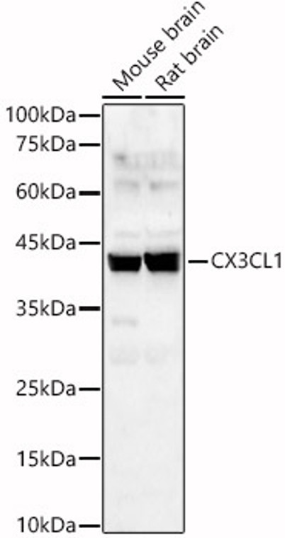
| Host: |
Rabbit |
| Applications: |
WB/IHC/IF |
| Reactivity: |
Human/Mouse/Rat |
| Note: |
STRICTLY FOR FURTHER SCIENTIFIC RESEARCH USE ONLY (RUO). MUST NOT TO BE USED IN DIAGNOSTIC OR THERAPEUTIC APPLICATIONS. |
| Short Description: |
Rabbit polyclonal antibody anti-CX3CL1 (365-397) is suitable for use in Western Blot, Immunohistochemistry and Immunofluorescence research applications. |
| Clonality: |
Polyclonal |
| Conjugation: |
Unconjugated |
| Isotype: |
IgG |
| Formulation: |
PBS with 0.01% Thimerosal, 50% Glycerol, pH7.3. |
| Purification: |
Affinity purification |
| Dilution Range: |
WB 1:500-1:1000IHC-P 1:50-1:200IF/ICC 1:50-1:200 |
| Storage Instruction: |
Store at-20°C for up to 1 year from the date of receipt, and avoid repeat freeze-thaw cycles. |
| Gene Symbol: |
CX3CL1 |
| Gene ID: |
6376 |
| Uniprot ID: |
X3CL1_HUMAN |
| Immunogen Region: |
365-397 |
| Immunogen: |
Recombinant fusion protein containing a sequence corresponding to amino acids 365-397 of human CX3CL1 (NP_002987.1). |
| Immunogen Sequence: |
LQGCPRKMAGEMAEGLRYIP RSCGSNSYVLVPV |
| Tissue Specificity | Expressed in the seminal plasma, endometrial fluid and follicular fluid (at protein level). Small intestine, colon, testis, prostate, heart, brain, lung, skeletal muscle, kidney and pancreas. Most abundant in the brain and heart. |
| Post Translational Modifications | A soluble short 95 kDa form may be released by proteolytic cleavage from the long membrane-anchored form. O-glycosylated with core 1 or possibly core 8 glycans. |
| Function | Chemokine that acts as a ligand for both CX3CR1 and integrins ITGAV:ITGB3 and ITGA4:ITGB1. The CX3CR1-CX3CL1 signaling exerts distinct functions in different tissue compartments, such as immune response, inflammation, cell adhesion and chemotaxis. Regulates leukocyte adhesion and migration processes at the endothelium. Can activate integrins in both a CX3CR1-dependent and CX3CR1-independent manner. In the presence of CX3CR1, activates integrins by binding to the classical ligand-binding site (site 1) in integrins. In the absence of CX3CR1, binds to a second site (site 2) in integrins which is distinct from site 1 and enhances the binding of other integrin ligands to site 1. Processed fractalkine: The soluble form is chemotactic for T-cells and monocytes, but not for neutrophils. Fractalkine: The membrane-bound form promotes adhesion of those leukocytes to endothelial cells. (Microbial infection) Mediates the cytoadherence of erythrocytes infected with parasite P.falciparum (strain 3D7) with endothelial cells by interacting with P.falciparum CBP1 and CBP2 expressed at the surface of erythrocytes. The adhesion prevents the elimination of infected erythrocytes by the spleen (Probable). |
| Protein Name | FractalkineC-X3-C Motif Chemokine 1Cx3c Membrane-Anchored ChemokineNeurotactinSmall-Inducible Cytokine D1 Cleaved Into - Processed Fractalkine |
| Database Links | Reactome: R-HSA-380108Reactome: R-HSA-418594 |
| Cellular Localisation | Cell MembraneSingle-Pass Type I Membrane ProteinProcessed Fractalkine: Secreted |
| Alternative Antibody Names | Anti-Fractalkine antibodyAnti-C-X3-C Motif Chemokine 1 antibodyAnti-Cx3c Membrane-Anchored Chemokine antibodyAnti-Neurotactin antibodyAnti-Small-Inducible Cytokine D1 Cleaved Into - Processed Fractalkine antibodyAnti-CX3CL1 antibodyAnti-FKN antibodyAnti-NTT antibodyAnti-SCYD1 antibodyAnti-A-152E5.2 antibody |
Information sourced from Uniprot.org
12 months for antibodies. 6 months for ELISA Kits. Please see website T&Cs for further guidance


![Immunofluorescence analysis of NIH/3T3 cells using [KO Validated] DNAJA1 Rabbit polyclonal antibody (STJ113207) at dilution of 1:100. Secondary antibody: Cy3 Goat Anti-Rabbit IgG (H+L) at 1:500 dilution. Blue: DAPI for nuclear staining. Immunofluorescence analysis of NIH/3T3 cells using [KO Validated] DNAJA1 Rabbit polyclonal antibody (STJ113207) at dilution of 1:100. Secondary antibody: Cy3 Goat Anti-Rabbit IgG (H+L) at 1:500 dilution. Blue: DAPI for nuclear staining.](https://cdn11.bigcommerce.com/s-zso2xnchw9/images/stencil/300x300/products/92785/373370/STJ113207_1__86638.1713139598.jpg?c=1)