| Host: |
Rabbit |
| Applications: |
WB/IHC/IF |
| Reactivity: |
Human/Mouse/Rat |
| Note: |
STRICTLY FOR FURTHER SCIENTIFIC RESEARCH USE ONLY (RUO). MUST NOT TO BE USED IN DIAGNOSTIC OR THERAPEUTIC APPLICATIONS. |
| Short Description: |
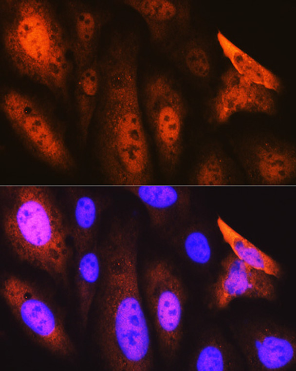
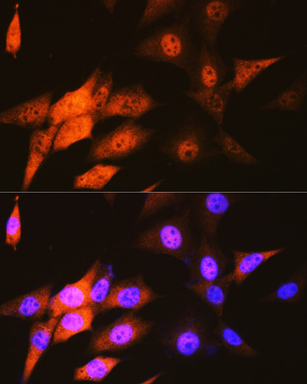
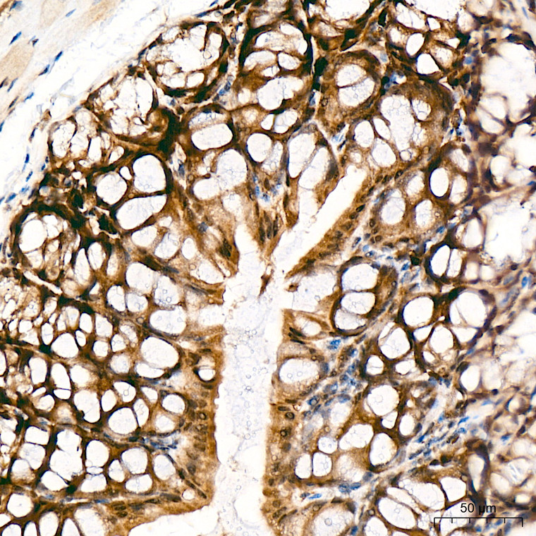
Rabbit monoclonal antibody anti-JAB1 (1-100) is suitable for use in Western Blot, Immunohistochemistry and Immunofluorescence research applications. |
| Clonality: |
Monoclonal |
| Clone ID: |
S3MR |
| Conjugation: |
Unconjugated |
| Isotype: |
IgG |
| Formulation: |
PBS with 0.02% Sodium Azide, 0.05% BSA, 50% Glycerol, pH7.3. |
| Purification: |
Affinity purification |
| Dilution Range: |
WB 1:500-1:2000IHC-P 1:50-1:200IF/ICC 1:50-1:200ChIP 1:50-1:200 |
| Storage Instruction: |
Store at-20°C for up to 1 year from the date of receipt, and avoid repeat freeze-thaw cycles. |
| Gene Symbol: |
COPS5 |
| Gene ID: |
10987 |
| Uniprot ID: |
CSN5_HUMAN |
| Immunogen Region: |
1-100 |
| Immunogen: |
Recombinant fusion protein containing a sequence corresponding to amino acids 1-100 of human JAB1/CSN5/COPS5 (Q92905). |
| Immunogen Sequence: |
MAASGSGMAQKTWELANNMQ EAQSIDEIYKYDKKQQQEIL AAKPWTKDHHYFKYCKISAL ALLKMVMHARSGGNLEVMGL MLGKVDGETMIIMDSFALPV |
| Function | Probable protease subunit of the COP9 signalosome complex (CSN), a complex involved in various cellular and developmental processes. The CSN complex is an essential regulator of the ubiquitin (Ubl) conjugation pathway by mediating the deneddylation of the cullin subunits of the SCF-type E3 ligase complexes, leading to decrease the Ubl ligase activity of SCF-type complexes such as SCF, CSA or DDB2. The complex is also involved in phosphorylation of p53/TP53, c-jun/JUN, IkappaBalpha/NFKBIA, ITPK1 and IRF8, possibly via its association with CK2 and PKD kinases. CSN-dependent phosphorylation of TP53 and JUN promotes and protects degradation by the Ubl system, respectively. In the complex, it probably acts as the catalytic center that mediates the cleavage of Nedd8 from cullins. It however has no metalloprotease activity by itself and requires the other subunits of the CSN complex. Interacts directly with a large number of proteins that are regulated by the CSN complex, confirming a key role in the complex. Promotes the proteasomal degradation of BRSK2. |
| Protein Name | Cop9 Signalosome Complex Subunit 5Sgn5Signalosome Subunit 5Jun Activation Domain-Binding Protein 1 |
| Database Links | Reactome: R-HSA-5696394Reactome: R-HSA-6781823Reactome: R-HSA-8856825Reactome: R-HSA-8951664 |
| Cellular Localisation | CytoplasmCytosolNucleusPerinuclear RegionCytoplasmic VesicleSecretory VesicleSynaptic VesicleNuclear Localization Is Diminished In The Presence Of Ifit3 |
| Alternative Antibody Names | Anti-Cop9 Signalosome Complex Subunit 5 antibodyAnti-Sgn5 antibodyAnti-Signalosome Subunit 5 antibodyAnti-Jun Activation Domain-Binding Protein 1 antibodyAnti-COPS5 antibodyAnti-CSN5 antibodyAnti-JAB1 antibody |
Information sourced from Uniprot.org
12 months for antibodies. 6 months for ELISA Kits. Please see website T&Cs for further guidance


