| Host: |
Rabbit |
| Applications: |
WB/IHC/IF |
| Reactivity: |
Human/Mouse/Rat |
| Note: |
STRICTLY FOR FURTHER SCIENTIFIC RESEARCH USE ONLY (RUO). MUST NOT TO BE USED IN DIAGNOSTIC OR THERAPEUTIC APPLICATIONS. |
| Short Description: |
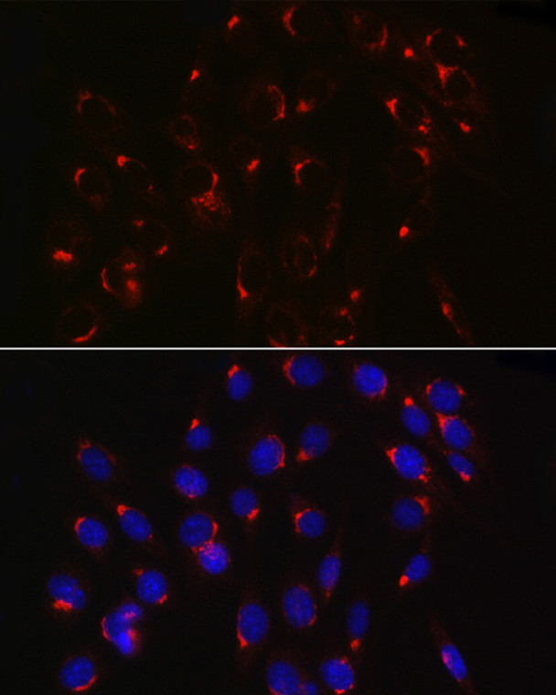
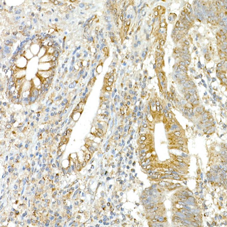
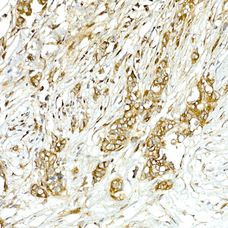
Rabbit polyclonal antibody anti-COPB2 (657-906) is suitable for use in Western Blot, Immunohistochemistry and Immunofluorescence research applications. |
| Clonality: |
Polyclonal |
| Conjugation: |
Unconjugated |
| Isotype: |
IgG |
| Formulation: |
PBS with 0.05% Proclin300, 50% Glycerol, pH7.3. |
| Purification: |
Affinity purification |
| Dilution Range: |
WB 1:100-1:500IHC-P 1:50-1:200IF/ICC 1:50-1:200 |
| Storage Instruction: |
Store at-20°C for up to 1 year from the date of receipt, and avoid repeat freeze-thaw cycles. |
| Gene Symbol: |
COPB2 |
| Gene ID: |
9276 |
| Uniprot ID: |
COPB2_HUMAN |
| Immunogen Region: |
657-906 |
| Immunogen: |
Recombinant fusion protein containing a sequence corresponding to amino acids 657-906 of human COPB2 (NP_004757.1). |
| Immunogen Sequence: |
AYQLAVEAESEQKWKQLAEL AISKCQFGLAQECLHHAQDY GGLLLLATASGNANMVNKLA EGAERDGKNNVAFMSYFLQG KVDACLELLIRTGRLPEAAF LARTYLPSQVSRVVKLWREN LSKVNQKAAESLADPTEYEN LFPGLKEAFVVEEWVKETHA DLWPAKQYPLVTPNEERNVM EEGKDFQPSRSTAQQELDGK PASPTPVIVASHTANKEEKS LLELEVDLDNLELEDIDTT |
| Function | The coatomer is a cytosolic protein complex that binds to dilysine motifs and reversibly associates with Golgi non-clathrin-coated vesicles, which further mediate biosynthetic protein transport from the ER, via the Golgi up to the trans Golgi network. Coatomer complex is required for budding from Golgi membranes, and is essential for the retrograde Golgi-to-ER transport of dilysine-tagged proteins. In mammals, the coatomer can only be recruited by membranes associated to ADP-ribosylation factors (ARFs), which are small GTP-binding proteins.the complex also influences the Golgi structural integrity, as well as the processing, activity, and endocytic recycling of LDL receptors. This coatomer complex protein, essential for Golgi budding and vesicular trafficking, is a selective binding protein (RACK) for protein kinase C, epsilon type. It binds to Golgi membranes in a GTP-dependent manner. |
| Protein Name | Coatomer Subunit Beta'Beta'-Coat ProteinBeta'-CopP102 |
| Database Links | Reactome: R-HSA-6807878Reactome: R-HSA-6811434 |
| Cellular Localisation | CytoplasmCytosolGolgi Apparatus MembranePeripheral Membrane ProteinCytoplasmic SideCytoplasmic VesicleCopi-Coated Vesicle MembraneThe Coatomer Is Cytoplasmic Or Polymerized On The Cytoplasmic Side Of The GolgiAs Well As On The Vesicles/Buds Originating From ItShows Only A Slight Preference For The Cis-Golgi ApparatusCompared With The Trans-Golgi |
| Alternative Antibody Names | Anti-Coatomer Subunit Beta' antibodyAnti-Beta'-Coat Protein antibodyAnti-Beta'-Cop antibodyAnti-P102 antibodyAnti-COPB2 antibody |
Information sourced from Uniprot.org
12 months for antibodies. 6 months for ELISA Kits. Please see website T&Cs for further guidance


