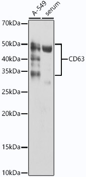
 at 1:1000 dilution. Secondary antibody: HRP Goat Anti-Rabbit IgG (H+L) (STJS000856) at 1:10000 dilution. Lysates/proteins: 25 Mu g per lane. Blocking buffer: 3% nonfat dry milk in TBST. Detection: ECL Basic Kit. Exposure time: 60s.")
 at 1:1000 dilution. Secondary antibody: HRP Goat Anti-Rabbit IgG (H+L) (STJS000856) at 1:10000 dilution. Lysates/proteins: 25 Mu g per lane. Blocking buffer: 3% nonfat dry milk in TBST. Detection: ECL Basic Kit. Exposure time: 60s.")
Anti-CD63 antibody (100-200) [S4602RM] (STJ11104602)
SPECIFICATIONS
ClonalityMonoclonal
HostRabbit
ConjugationUnconjugated
IsotypeIgG
General Information
| Short Description | Rabbit monoclonal Cd63 antigen (100-200) antibody for use in WB, IHC-P and ELISA in human, mouse and rat samples. Datasheet included with dilution recommendations, and related reagents. |
| Applications | WB/IHC-P/ELISA |
| Host | Rabbit |
| Reactivity | Human/Mouse/Rat |
| Note | STRICTLY FOR FURTHER SCIENTIFIC RESEARCH USE ONLY (RUO). MUST NOT TO BE USED IN DIAGNOSTIC OR THERAPEUTIC APPLICATIONS. |
Product Properties
| Clonality | Monoclonal |
| Clone ID | S4602RM |
| Isotype | IgG |
| Conjugation | Unconjugated |
| Concentration | Lot specific |
| Purification | Affinity purification |
| Dilution Range | WB:1:500-1:1000IHC-P:1:50-1:200ELISA:Recommended starting concentration is 1 Mu g/mL. Please optimize the concentration based on your specific assay requirements. |
| Formulation | PBS with 0.02% Sodium Azide, 0.05% BSA, 50% Glycerol, pH 7.3. |
| Storage Instruction | Store at-20°C for up to 1 year from the date of receipt, and avoid repeat freeze-thaw cycles. |
Target Information
| Gene Symbol | CD63 |
| Gene ID | 967 |
| Uniprot ID | CD63_HUMAN |
| Immunogen Region | 100-200 |
| Immunogen Sequence | AAIAGYVFRDKVMSEFNNNF RQQMENYPKNNHTASILDRM QADFKCCGAANYTDWEKIPS MSKNRVPDSCCINVTVGCGI NFNEKAIHKEGCVEKIGGWL R |
| Specificity | A synthetic peptide corresponding to a sequence within amino acids 100-200 of human CD63 (P08962). |
Additional Info
| Tissue Specificity | Detected in platelets (at protein level). Dysplastic nevi, radial growth phase primary melanomas, hematopoietic cells, tissue macrophages. |
| Post Translational Modifications | Palmitoylated at a low, basal level in unstimulated platelets. The level of palmitoylation increases when platelets are activated by thrombin (in vitro). |
| Function | Functions as a cell surface receptor for TIMP1 and plays a role in the activation of cellular signaling cascades. Plays a role in the activation of ITGB1 and integrin signaling, leading to the activation of AKT, FAK/PTK2 and MAP kinases. Promotes cell survival, reorganization of the actin cytoskeleton, cell adhesion, spreading and migration, via its role in the activation of AKT and FAK/PTK2. Plays a role in VEGFA signaling via its role in regulating the internalization of KDR/VEGFR2. Plays a role in intracellular vesicular transport processes, and is required for normal trafficking of the PMEL luminal domain that is essential for the development and maturation of melanocytes. Plays a role in the adhesion of leukocytes onto endothelial cells via its role in the regulation of SELP trafficking. May play a role in mast cell degranulation in response to Ms4a2/FceRI stimulation, but not in mast cell degranulation in response to other stimuli. |
| Protein Name | Cd63 AntigenGranulophysinLysosomal-Associated Membrane Protein 3Lamp-3Lysosome Integral Membrane Protein 1Limp1Melanoma-Associated Antigen Me491Oma81hOcular Melanoma-Associated AntigenTetraspanin-30Tspan-30Cd Antigen Cd63 |
| Database Links | Reactome: R-HSA-114608Reactome: R-HSA-6798695 |
| Cellular Localisation | Cell MembraneMulti-Pass Membrane ProteinLysosome MembraneLate Endosome MembraneEndosomeMultivesicular BodyMelanosomeSecretedExtracellular ExosomeCell SurfaceAlso Found In Weibel-Palade Bodies Of Endothelial CellsLocated In Platelet Dense GranulesDetected In A Subset Of Pre-MelanosomesDetected On Intralumenal Vesicles (Ilvs) Within Multivesicular Bodies |
| Alternative Antibody Names | Anti-Cd63 Antigen antibodyAnti-Granulophysin antibodyAnti-Lysosomal-Associated Membrane Protein 3 antibodyAnti-Lamp-3 antibodyAnti-Lysosome Integral Membrane Protein 1 antibodyAnti-Limp1 antibodyAnti-Melanoma-Associated Antigen Me491 antibodyAnti-Oma81h antibodyAnti-Ocular Melanoma-Associated Antigen antibodyAnti-Tetraspanin-30 antibodyAnti-Tspan-30 antibodyAnti-Cd Antigen Cd63 antibodyAnti-CD63 antibodyAnti-MLA1 antibodyAnti-TSPAN30 antibody |
Information sourced from Uniprot.org