| Host: |
Rabbit |
| Applications: |
WB/IF |
| Reactivity: |
Human/Mouse |
| Note: |
STRICTLY FOR FURTHER SCIENTIFIC RESEARCH USE ONLY (RUO). MUST NOT TO BE USED IN DIAGNOSTIC OR THERAPEUTIC APPLICATIONS. |
| Short Description: |
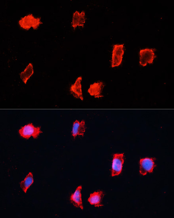
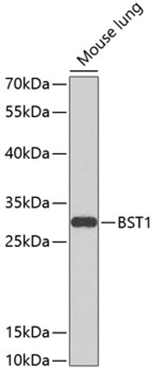
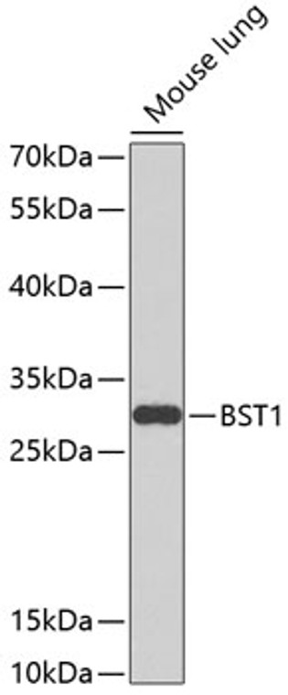
Rabbit polyclonal antibody anti-BST1 (29-293) is suitable for use in Western Blot and Immunofluorescence research applications. |
| Clonality: |
Polyclonal |
| Conjugation: |
Unconjugated |
| Isotype: |
IgG |
| Formulation: |
PBS with 0.02% Sodium Azide, 50% Glycerol, pH7.3. |
| Purification: |
Affinity purification |
| Dilution Range: |
WB 1:500-1:2000IF/ICC 1:50-1:200 |
| Storage Instruction: |
Store at-20°C for up to 1 year from the date of receipt, and avoid repeat freeze-thaw cycles. |
| Gene Symbol: |
BST1 |
| Gene ID: |
683 |
| Uniprot ID: |
BST1_HUMAN |
| Immunogen Region: |
29-293 |
| Immunogen: |
Recombinant fusion protein containing a sequence corresponding to amino acids 29-293 of human BST1 (NP_004325.2). |
| Immunogen Sequence: |
GARARWRGEGTSAHLRDIFL GRCAEYRALLSPEQRNKNCT AIWEAFKVALDKDPCSVLPS DYDLFINLSRHSIPRDKSLF WENSHLLVNSFADNTRRFMP LSDVLYGRVADFLSWCRQKN DSGLDYQSCPTSEDCENNPV DSFWKRASIQYSKDSSGVIH VMLNGSEPTGAYPIKGFFAD YEIPNLQKEKITRIEIWVMH EIGGPNVESCGEGSMKVLEK RLKDMGFQYSCINDYRPVK |
| Tissue Specificity | Expressed in various tissues including placenta, lung, liver and kidney. |
| Function | Catalyzes both the synthesis of cyclic ADP-beta-D-ribose (cADPR) from NAD(+), and its hydrolysis to ADP-D-ribose (ADPR). Cyclic ADPR is known to serve as an endogenous second messenger that elicits calcium release from intracellular stores, and thus regulates the mobilization of intracellular calcium (Probable). May be involved in pre-B-cell growth (Probable). |
| Protein Name | Adp-Ribosyl Cyclase/Cyclic Adp-Ribose Hydrolase 2Adp-Ribosyl Cyclase 2Bone Marrow Stromal Cell Antigen 1Bst-1Cyclic Adp-Ribose Hydrolase 2Cadpr Hydrolase 2Cd Antigen Cd157 |
| Database Links | Reactome: R-HSA-163125Reactome: R-HSA-196807Reactome: R-HSA-6798695 |
| Cellular Localisation | Cell MembraneLipid-AnchorGpi-Anchor |
| Alternative Antibody Names | Anti-Adp-Ribosyl Cyclase/Cyclic Adp-Ribose Hydrolase 2 antibodyAnti-Adp-Ribosyl Cyclase 2 antibodyAnti-Bone Marrow Stromal Cell Antigen 1 antibodyAnti-Bst-1 antibodyAnti-Cyclic Adp-Ribose Hydrolase 2 antibodyAnti-Cadpr Hydrolase 2 antibodyAnti-Cd Antigen Cd157 antibodyAnti-BST1 antibody |
Information sourced from Uniprot.org
12 months for antibodies. 6 months for ELISA Kits. Please see website T&Cs for further guidance


![Western blot analysis of lysates from wild type (WT) and MPG knockout (KO) HeLa cells, using [KO Validated] MPG Rabbit polyclonal antibody (STJ11100043) at 1:1000 dilution. Secondary antibody: HRP Goat Anti-Rabbit IgG (H+L) (STJS000856) at 1:10000 dilution. Lysates/proteins: 25 Mu g per lane. Blocking buffer: 3% nonfat dry milk in TBST. Detection: ECL Basic Kit. Exposure time: 60s. Western blot analysis of lysates from wild type (WT) and MPG knockout (KO) HeLa cells, using [KO Validated] MPG Rabbit polyclonal antibody (STJ11100043) at 1:1000 dilution. Secondary antibody: HRP Goat Anti-Rabbit IgG (H+L) (STJS000856) at 1:10000 dilution. Lysates/proteins: 25 Mu g per lane. Blocking buffer: 3% nonfat dry milk in TBST. Detection: ECL Basic Kit. Exposure time: 60s.](https://cdn11.bigcommerce.com/s-zso2xnchw9/images/stencil/300x300/products/89115/357567/STJ11100043_1__91960.1713121694.jpg?c=1)