• Immunohistochemistry analysis of paraffin-embedded human breast carcinoma tissue, using Survivin Antibody. The picture on the right is blocked with the synthesized peptide.
  • Western blot analysis of lysates from mouse lung, using Survivin Antibody. The lane on the right is blocked with the synthesized peptide.
  • Immunohistochemical analysis of paraffin-embedded Mouse-kidney tissue. 1, Survivin Polyclonal Antibody was diluted at 1:200 (4°C, overnight). 2, Sodium citrate pH 6.0 was used for antibody retrieval (>98°C, 20min). 3, Secondary antibody was diluted at 1:200 (room tempeRature, 30min). Negative control was used by secondary antibody only.
  • Immunofluorescence analysis of HeLa cells, using Survivin Antibody. The picture on the right is blocked with the synthesized peptide.
  • Immunohistochemical analysis of paraffin-embedded Rat-spleen tissue. 1, Survivin Polyclonal Antibody was diluted at 1:200 (4°C, overnight). 2, Sodium citrate pH 6.0 was used for antibody retrieval (>98°C, 20min). 3, Secondary antibody was diluted at 1:200 (room tempeRature, 30min). Negative control was used by secondary antibody only.
  • Immunohistochemical analysis of paraffin-embedded Mouse-testis tissue. 1, Survivin Polyclonal Antibody was diluted at 1:200 (4°C, overnight). 2, Sodium citrate pH 6.0 was used for antibody retrieval (>98°C, 20min). 3, Secondary antibody was diluted at 1:200 (room tempeRature, 30min). Negative control was used by secondary antibody only.
  • Immunohistochemical analysis of paraffin-embedded Human-lung-cancer tissue. 1, Survivin Polyclonal Antibody was diluted at 1:200 (4°C, overnight). 2, Sodium citrate pH 6.0 was used for antibody retrieval (>98°C, 20min). 3, Secondary antibody was diluted at 1:200 (room tempeRature, 30min). Negative control was used by secondary antibody only.
  • Immunohistochemical analysis of paraffin-embedded Rat-testis tissue. 1, Survivin Polyclonal Antibody was diluted at 1:200 (4°C, overnight). 2, Sodium citrate pH 6.0 was used for antibody retrieval (>98°C, 20min). 3, Secondary antibody was diluted at 1:200 (room tempeRature, 30min). Negative control was used by secondary antibody only.
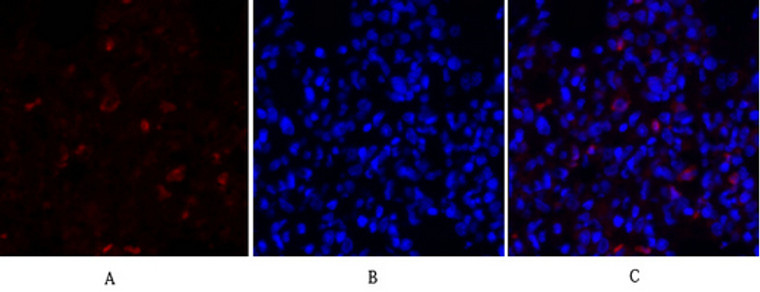
  • Immunofluorescence analysis of mouse-lung tissue. 1, Survivin Polyclonal Antibody (red) was diluted at 1:200 (4°C, overnight). 2, Cy3 labled Secondary antibody was diluted at 1:300 (room temperature, 50min).3, Picture B: DAPI (blue) 10min. Picture A:Target. Picture B: DAPI. Picture C: merge of A+B
  • Immunofluorescence analysis of mouse-lung tissue. 1, Survivin Polyclonal Antibody (red) was diluted at 1:200 (4°C, overnight). 2, Cy3 labled Secondary antibody was diluted at 1:300 (room temperature, 50min).3, Picture B: DAPI (blue) 10min. Picture A:Target. Picture B: DAPI. Picture C: merge of A+B
  • Immunofluorescence analysis of rat-spleen tissue. 1, Survivin Polyclonal Antibody (red) was diluted at 1:200 (4°C, overnight). 2, Cy3 labled Secondary antibody was diluted at 1:300 (room temperature, 50min).3, Picture B: DAPI (blue) 10min. Picture A:Target. Picture B: DAPI. Picture C: merge of A+B
  • Immunofluorescence analysis of rat-spleen tissue. 1, Survivin Polyclonal Antibody (red) was diluted at 1:200 (4°C, overnight). 2, Cy3 labled Secondary antibody was diluted at 1:300 (room temperature, 50min).3, Picture B: DAPI (blue) 10min. Picture A:Target. Picture B: DAPI. Picture C: merge of A+B
  • Immunofluorescence analysis of rat-lung tissue. 1, Survivin Polyclonal Antibody (red) was diluted at 1:200 (4°C, overnight). 2, Cy3 labled Secondary antibody was diluted at 1:300 (room temperature, 50min).3, Picture B: DAPI (blue) 10min. Picture A:Target. Picture B: DAPI. Picture C: merge of A+B
  • Immunofluorescence analysis of rat-lung tissue. 1, Survivin Polyclonal Antibody (red) was diluted at 1:200 (4°C, overnight). 2, Cy3 labled Secondary antibody was diluted at 1:300 (room temperature, 50min).3, Picture B: DAPI (blue) 10min. Picture A:Target. Picture B: DAPI. Picture C: merge of A+B
  • Immunofluorescence analysis of Hela cell. 1, Survivin Polyclonal Antibody (red) was diluted at 1:200 (4°C overnight). Active Caspase-3 monoclonal antibody (5E1) (green) was diluted at 1:200 (4°C overnight). 2, Goat Anti Rabbit Alexa Fluor 594 Catalog: (NA was diluted at 1:1000 (room temperature, 50min). Goat Anti Mouse Alexa Fluor 488 Catalog: (NA was diluted at 1:1000 (room temperature, 50min).

Anti-BIRC5 antibody (86-135 aa) (STJ95838)

SKU:
STJ95838

Current Stock:
Host: Rabbit
Applications: WB/IHC/IF/ELISA
Reactivity: Human/Mouse/Rat
Note: STRICTLY FOR FURTHER SCIENTIFIC RESEARCH USE ONLY (RUO). MUST NOT TO BE USED IN DIAGNOSTIC OR THERAPEUTIC APPLICATIONS.
Short Description: Rabbit polyclonal antibody anti-Baculoviral IAP repeat-containing protein 5 (86-135 aa) is suitable for use in Western Blot, Immunohistochemistry, Immunofluorescence and ELISA research applications.
Clonality: Polyclonal
Conjugation: Unconjugated
Isotype: IgG
Formulation: Liquid in PBS containing 50% Glycerol, 0.5% BSA and 0.02% Sodium Azide.
Purification: The antibody was affinity-purified from rabbit antiserum by affinity-chromatography using epitope-specific immunogen.
Concentration: 1 mg/mL
Dilution Range: WB 1:500-1:2000
IHC 1:100-1:300
IF 1:200-1:1000
ELISA 1:5000
Storage Instruction: Store at-20°C for up to 1 year from the date of receipt, and avoid repeat freeze-thaw cycles.
Gene Symbol: BIRC5
Gene ID: 332
Uniprot ID: BIRC5_HUMAN
Immunogen Region: 86-135 aa
Specificity: Survivin Polyclonal Antibody detects endogenous levels of Survivin protein.
Immunogen: The antiserum was produced against synthesized peptide derived from the human Survivin at the amino acid range 86-135
Post Translational Modifications Ubiquitinated by the Cul9-RING ubiquitin-protein ligase complex, leading to its degradation. Ubiquitination is required for centrosomal targeting. Deubiquitinated by USP35 or USP38.leading to stabilization. In vitro phosphorylation at Thr-117 by AURKB prevents interaction with INCENP and localization to mitotic chromosomes. Phosphorylation at Thr-48 by CK2 is critical for its mitotic and anti-apoptotic activities. Phosphorylation at Thr-34 by CDK15 is critical for its anti-apoptotic activity. Phosphorylation at Ser-20 by AURKC is critical for regulation of proper chromosome alignment and segregation, and possibly cytokinesis. Acetylation at Lys-129 by CBP results in its homodimerization, while deacetylation promotes the formation of monomers which heterodimerize with XPO1/CRM1 which facilitates its nuclear export. The acetylated form represses STAT3 transactivation. The dynamic equilibrium between its acetylation and deacetylation at Lys-129 determines its interaction with XPO1/CRM1, its subsequent subcellular localization, and its ability to inhibit STAT3 transactivation.
Function Multitasking protein that has dual roles in promoting cell proliferation and preventing apoptosis. Component of a chromosome passage protein complex (CPC) which is essential for chromosome alignment and segregation during mitosis and cytokinesis. Acts as an important regulator of the localization of this complex.directs CPC movement to different locations from the inner centromere during prometaphase to midbody during cytokinesis and participates in the organization of the center spindle by associating with polymerized microtubules. Involved in the recruitment of CPC to centromeres during early mitosis via association with histone H3 phosphorylated at 'Thr-3' (H3pT3) during mitosis. The complex with RAN plays a role in mitotic spindle formation by serving as a physical scaffold to help deliver the RAN effector molecule TPX2 to microtubules. May counteract a default induction of apoptosis in G2/M phase. The acetylated form represses STAT3 transactivation of target gene promoters. May play a role in neoplasia. Inhibitor of CASP3 and CASP7. Essential for the maintenance of mitochondrial integrity and function. Isoform 2 and isoform 3 do not appear to play vital roles in mitosis. Isoform 3 shows a marked reduction in its anti-apoptotic effects when compared with the displayed wild-type isoform.
Protein Name Baculoviral Iap Repeat-Containing Protein 5
Apoptosis Inhibitor 4
Apoptosis Inhibitor Survivin
Database Links Reactome: R-HSA-141444
Reactome: R-HSA-2467813
Reactome: R-HSA-2500257
Reactome: R-HSA-4615885
Reactome: R-HSA-5663220
Reactome: R-HSA-6785807
Reactome: R-HSA-6803205
Reactome: R-HSA-68877
Reactome: R-HSA-8951664
Reactome: R-HSA-9648025
Cellular Localisation Cytoplasm
Nucleus
Chromosome
Centromere
Cytoskeleton
Spindle
Kinetochore
Midbody
Localizes At The Centromeres From Prophase To Metaphase
At The Spindle Midzone During Anaphase And A The Midbody During Telophase And Cytokinesis
Accumulates In The Nucleus Upon Treatment With Leptomycin B (Lmb)
A Xpo1/Crm1 Nuclear Export Inhibitor
Localizes On Chromosome Arms And Inner Centromeres From Prophase Through Metaphase
Localizes To Kinetochores In Metaphase
Distributes To The Midzone Microtubules In Anaphase And At Telophase
Localizes Exclusively To The Midbody
Colocalizes With Aurkb At Mitotic Chromosomes
Acetylation At Lys-129 Directs Its Localization To The Nucleus By Enhancing Homodimerization And Thereby Inhibiting Xpo1/Crm1-Mediated Nuclear Export
Alternative Antibody Names Anti-Baculoviral Iap Repeat-Containing Protein 5 antibody
Anti-Apoptosis Inhibitor 4 antibody
Anti-Apoptosis Inhibitor Survivin antibody
Anti-BIRC5 antibody
Anti-API4 antibody
Anti-IAP4 antibody

Information sourced from Uniprot.org

12 months for antibodies. 6 months for ELISA Kits. Please see website T&Cs for further guidance