| Host: |
Rabbit |
| Applications: |
WB/IHC |
| Reactivity: |
Human/Mouse/Rat |
| Note: |
STRICTLY FOR FURTHER SCIENTIFIC RESEARCH USE ONLY (RUO). MUST NOT TO BE USED IN DIAGNOSTIC OR THERAPEUTIC APPLICATIONS. |
| Short Description: |
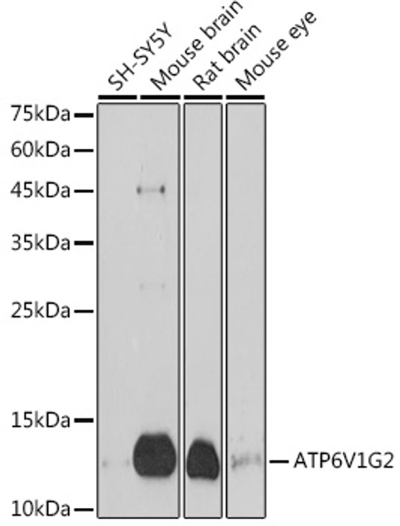
Rabbit polyclonal antibody anti-ATP6V1G2 (40-100) is suitable for use in Western Blot and Immunohistochemistry research applications. |
| Clonality: |
Polyclonal |
| Conjugation: |
Unconjugated |
| Isotype: |
IgG |
| Formulation: |
PBS with 0.01% Thimerosal, 50% Glycerol, pH7.3. |
| Purification: |
Affinity purification |
| Dilution Range: |
WB 1:500-1:1000IHC-P 1:50-1:200 |
| Storage Instruction: |
Store at-20°C for up to 1 year from the date of receipt, and avoid repeat freeze-thaw cycles. |
| Gene Symbol: |
ATP6V1G2 |
| Gene ID: |
534 |
| Uniprot ID: |
VATG2_HUMAN |
| Immunogen Region: |
40-100 |
| Immunogen: |
Recombinant fusion protein containing a sequence corresponding to amino acids 40-100 of human ATP6V1G2 (NP_569730.1). |
| Immunogen Sequence: |
AQMEVEQYRREREHEFQSKQ QAAMGSQGNLSAEVEQATRR QVQGMQSSQQRNRERVLAQL L |
| Tissue Specificity | Brain. |
| Function | Subunit of the V1 complex of vacuolar(H+)-ATPase (V-ATPase), a multisubunit enzyme composed of a peripheral complex (V1) that hydrolyzes ATP and a membrane integral complex (V0) that translocates protons. V-ATPase is responsible for acidifying and maintaining the pH of intracellular compartments and in some cell types, is targeted to the plasma membrane, where it is responsible for acidifying the extracellular environment. |
| Protein Name | V-Type Proton Atpase Subunit G 2V-Atpase Subunit G 2V-Atpase 13 Kda Subunit 2Vacuolar Proton Pump Subunit G 2 |
| Database Links | Reactome: R-HSA-1222556Reactome: R-HSA-77387Reactome: R-HSA-917977Reactome: R-HSA-9639288Reactome: R-HSA-983712 |
| Cellular Localisation | MelanosomeCytoplasmic VesicleClathrin-Coated Vesicle MembranePeripheral Membrane ProteinHighly Enriched In Late-Stage Melanosomes |
| Alternative Antibody Names | Anti-V-Type Proton Atpase Subunit G 2 antibodyAnti-V-Atpase Subunit G 2 antibodyAnti-V-Atpase 13 Kda Subunit 2 antibodyAnti-Vacuolar Proton Pump Subunit G 2 antibodyAnti-ATP6V1G2 antibodyAnti-ATP6G antibodyAnti-ATP6G2 antibodyAnti-NG38 antibody |
Information sourced from Uniprot.org
12 months for antibodies. 6 months for ELISA Kits. Please see website T&Cs for further guidance


