| Host: |
Rabbit |
| Applications: |
WB/IF/ELISA |
| Reactivity: |
Human/Mouse |
| Note: |
STRICTLY FOR FURTHER SCIENTIFIC RESEARCH USE ONLY (RUO). MUST NOT TO BE USED IN DIAGNOSTIC OR THERAPEUTIC APPLICATIONS. |
| Short Description: |
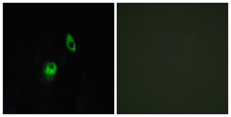
Rabbit polyclonal antibody anti-Adhesion G protein-coupled receptor A2 (1151-1200 aa) is suitable for use in Western Blot, Immunofluorescence and ELISA research applications. |
| Clonality: |
Polyclonal |
| Conjugation: |
Unconjugated |
| Isotype: |
IgG |
| Formulation: |
Liquid in PBS containing 50% Glycerol, 0.5% BSA and 0.02% Sodium Azide. |
| Purification: |
The antibody was affinity-purified from rabbit antiserum by affinity-chromatography using epitope-specific immunogen. |
| Concentration: |
1 mg/mL |
| Dilution Range: |
WB 1:500-1:2000IF 1:200-1:1000ELISA 1:10000 |
| Storage Instruction: |
Store at-20°C for up to 1 year from the date of receipt, and avoid repeat freeze-thaw cycles. |
| Gene Symbol: |
ADGRA2 |
| Gene ID: |
25960 |
| Uniprot ID: |
AGRA2_HUMAN |
| Immunogen Region: |
1151-1200 aa |
| Specificity: |
GPR124 Polyclonal Antibody detects endogenous levels of GPR124 protein. |
| Immunogen: |
The antiserum was produced against synthesized peptide derived from the human GPR124 at the amino acid range 1151-1200 |
| Post Translational Modifications | Glycosylated. Proteolytically cleaved into two subunits, an extracellular subunit and a seven-transmembrane subunit. Cleaved by thrombin (F2) and MMP1. Also cleaved by MMP9, with lower efficiency. Presence of the protein disulfide-isomerase P4HB at the cell surface is additionally required for shedding of the extracellular subunit, suggesting that the subunits are linked by disulfide bonds. Shedding is enhanced by the growth factor FGF2 and may promote cell survival during angiogenesis. |
| Function | Endothelial receptor which functions together with RECK to enable brain endothelial cells to selectively respond to Wnt7 signals (WNT7A or WNT7B). Plays a key role in Wnt7-specific responses, such as endothelial cell sprouting and migration in the forebrain and neural tube, and establishment of the blood-brain barrier. Acts as a Wnt7-specific coactivator of canonical Wnt signaling: required to deliver RECK-bound Wnt7 to frizzled by assembling a higher-order RECK-ADGRA2-Fzd-LRP5-LRP6 complex. ADGRA2-tethering function does not rely on its G-protein coupled receptor (GPCR) structure but instead on its combined capacity to interact with RECK extracellularly and recruit the Dishevelled scaffolding protein intracellularly. Binds to the glycosaminoglycans heparin, heparin sulfate, chondroitin sulfate and dermatan sulfate. |
| Protein Name | Adhesion G Protein-Coupled Receptor A2G-Protein Coupled Receptor 124Tumor Endothelial Marker 5 |
| Cellular Localisation | Cell MembraneMulti-Pass Membrane ProteinCell ProjectionFilopodiumEnriched At Lateral Cell Borders And Also At Sites Of Cell-Ecm (Extracellular Matrix) Contact |
| Alternative Antibody Names | Anti-Adhesion G Protein-Coupled Receptor A2 antibodyAnti-G-Protein Coupled Receptor 124 antibodyAnti-Tumor Endothelial Marker 5 antibodyAnti-ADGRA2 antibodyAnti-GPR124 antibodyAnti-KIAA1531 antibodyAnti-TEM5 antibody |
Information sourced from Uniprot.org
12 months for antibodies. 6 months for ELISA Kits. Please see website T&Cs for further guidance


