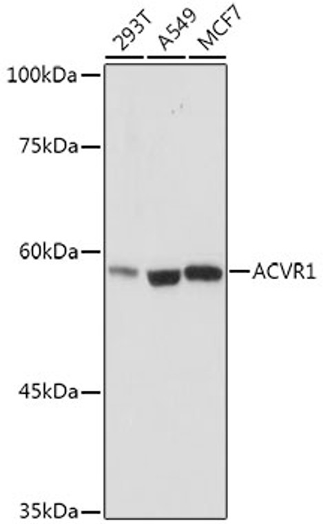
| Host: |
Rabbit |
| Applications: |
WB |
| Reactivity: |
Human/Mouse/Rat |
| Note: |
STRICTLY FOR FURTHER SCIENTIFIC RESEARCH USE ONLY (RUO). MUST NOT TO BE USED IN DIAGNOSTIC OR THERAPEUTIC APPLICATIONS. |
| Short Description: |
Rabbit monoclonal antibody anti-ACVR1 (410-509) is suitable for use in Western Blot research applications. |
| Clonality: |
Monoclonal |
| Clone ID: |
S5MR |
| Conjugation: |
Unconjugated |
| Isotype: |
IgG |
| Formulation: |
PBS with 0.02% Sodium Azide, 0.05% BSA, 50% Glycerol, pH7.3. |
| Purification: |
Affinity purification |
| Dilution Range: |
WB 1:500-1:1000 |
| Storage Instruction: |
Store at-20°C for up to 1 year from the date of receipt, and avoid repeat freeze-thaw cycles. |
| Gene Symbol: |
ACVR1 |
| Gene ID: |
90 |
| Uniprot ID: |
ACVR1_HUMAN |
| Immunogen Region: |
410-509 |
| Immunogen: |
A synthetic peptide corresponding to a sequence within amino acids 410-509 of human ACVR1 (Q04771). |
| Immunogen Sequence: |
VLWEVARRMVSNGIVEDYKP PFYDVVPNDPSFEDMRKVVC VDQQRPNIPNRWFSDPTLTS LAKLMKECWYQNPSARLTAL RIKKTLTKIDNSLDKLKTDC |
| Tissue Specificity | Expressed in normal parenchymal cells, endothelial cells, fibroblasts and tumor-derived epithelial cells. |
| Function | Bone morphogenetic protein (BMP) type I receptor that is involved in a wide variety of biological processes, including bone, heart, cartilage, nervous, and reproductive system development and regulation. As a type I receptor, forms heterotetrameric receptor complexes with the type II receptors AMHR2, ACVR2A or ACVR2B. Upon binding of ligands such as BMP7 or GDF2/BMP9 to the heteromeric complexes, type II receptors transphosphorylate ACVR1 intracellular domain. In turn, ACVR1 kinase domain is activated and subsequently phosphorylates SMAD1/5/8 proteins that transduce the signal. In addition to its role in mediating BMP pathway-specific signaling, suppresses TGFbeta/activin pathway signaling by interfering with the binding of activin to its type II receptor. Besides canonical SMAD signaling, can activate non-canonical pathways such as p38 mitogen-activated protein kinases/MAPKs. May promote the expression of HAMP, potentially via its interaction with BMP6. |
| Protein Name | Activin Receptor Type-1Activin Receptor Type IActr-IActivin Receptor-Like Kinase 2Alk-2Serine/Threonine-Protein Kinase Receptor R1Skr1Tgf-B Superfamily Receptor Type ITsr-I |
| Cellular Localisation | MembraneSingle-Pass Type I Membrane Protein |
| Alternative Antibody Names | Anti-Activin Receptor Type-1 antibodyAnti-Activin Receptor Type I antibodyAnti-Actr-I antibodyAnti-Activin Receptor-Like Kinase 2 antibodyAnti-Alk-2 antibodyAnti-Serine/Threonine-Protein Kinase Receptor R1 antibodyAnti-Skr1 antibodyAnti-Tgf-B Superfamily Receptor Type I antibodyAnti-Tsr-I antibodyAnti-ACVR1 antibodyAnti-ACVRLK2 antibody |
Information sourced from Uniprot.org
12 months for antibodies. 6 months for ELISA Kits. Please see website T&Cs for further guidance


