| Host: |
Rabbit |
| Applications: |
WB/IHC/IF/ELISA |
| Reactivity: |
Human/Rat/Mouse |
| Note: |
STRICTLY FOR FURTHER SCIENTIFIC RESEARCH USE ONLY (RUO). MUST NOT TO BE USED IN DIAGNOSTIC OR THERAPEUTIC APPLICATIONS. |
| Short Description: |
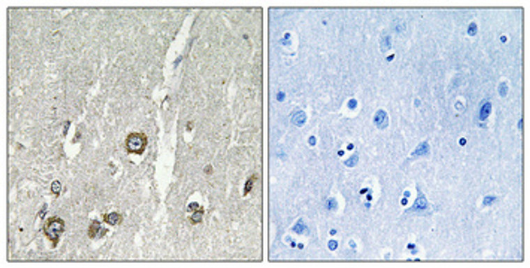
Rabbit polyclonal antibody anti-Acyl-coenzyme A thioesterase 8 (131-180 aa) is suitable for use in Western Blot, Immunohistochemistry, Immunofluorescence and ELISA research applications. |
| Clonality: |
Polyclonal |
| Conjugation: |
Unconjugated |
| Isotype: |
IgG |
| Formulation: |
Liquid in PBS containing 50% Glycerol, 0.5% BSA and 0.02% Sodium Azide. |
| Purification: |
The antibody was affinity-purified from rabbit antiserum by affinity-chromatography using epitope-specific immunogen. |
| Concentration: |
1 mg/mL |
| Dilution Range: |
WB 1:500-1:2000IHC 1:100-1:300IF 1:200-1:1000ELISA 1:40000 |
| Storage Instruction: |
Store at-20°C for up to 1 year from the date of receipt, and avoid repeat freeze-thaw cycles. |
| Gene Symbol: |
ACOT8 |
| Gene ID: |
10005 |
| Uniprot ID: |
ACOT8_HUMAN |
| Immunogen Region: |
131-180 aa |
| Specificity: |
ACOT8 Polyclonal Antibody detects endogenous levels of ACOT8 protein. |
| Immunogen: |
The antiserum was produced against synthesized peptide derived from the human ACOT8 at the amino acid range 131-180 |
| Function | Catalyzes the hydrolysis of acyl-CoAs into free fatty acids and coenzyme A (CoASH), regulating their respective intracellular levels. Displays no strong substrate specificity with respect to the carboxylic acid moiety of Acyl-CoAs. Hydrolyzes medium length (C2 to C20) straight-chain, saturated and unsaturated acyl-CoAS but is inactive towards substrates with longer aliphatic chains. Moreover, it catalyzes the hydrolysis of CoA esters of bile acids, such as choloyl-CoA and chenodeoxycholoyl-CoA and competes with bile acid CoA:amino acid N-acyltransferase (BAAT). Is also able to hydrolyze CoA esters of dicarboxylic acids. It is involved in the metabolic regulation of peroxisome proliferation. (Microbial infection) May mediate Nef-induced down-regulation of CD4 cell-surface expression. |
| Protein Name | Acyl-Coenzyme A Thioesterase 8Acyl-Coa Thioesterase 8 Choloyl-Coenzyme A ThioesteraseHiv-Nef-Associated Acyl-Coa ThioesterasePeroxisomal Acyl-Coa Thioesterase 2Pte-2Peroxisomal Acyl-Coenzyme A Thioester Hydrolase 1Pte-1Peroxisomal Long-Chain Acyl-Coa Thioesterase 1Thioesterase IiHacte-IiiHacteiiiHte |
| Database Links | Reactome: R-HSA-193368Reactome: R-HSA-2046106Reactome: R-HSA-389887Reactome: R-HSA-390247Reactome: R-HSA-9033241 |
| Cellular Localisation | Peroxisome MatrixPredominantly Localized In The Peroxisome But A Localization To The Cytosol Cannot Be Excluded |
| Alternative Antibody Names | Anti-Acyl-Coenzyme A Thioesterase 8 antibodyAnti-Acyl-Coa Thioesterase 8 antibodyAnti-Choloyl-Coenzyme A Thioesterase antibodyAnti-Hiv-Nef-Associated Acyl-Coa Thioesterase antibodyAnti-Peroxisomal Acyl-Coa Thioesterase 2 antibodyAnti-Pte-2 antibodyAnti-Peroxisomal Acyl-Coenzyme A Thioester Hydrolase 1 antibodyAnti-Pte-1 antibodyAnti-Peroxisomal Long-Chain Acyl-Coa Thioesterase 1 antibodyAnti-Thioesterase Ii antibodyAnti-Hacte-Iii antibodyAnti-Hacteiii antibodyAnti-Hte antibodyAnti-ACOT8 antibodyAnti-ACTEIII antibodyAnti-PTE1 antibody |
Information sourced from Uniprot.org
12 months for antibodies. 6 months for ELISA Kits. Please see website T&Cs for further guidance


