-
CUT&Tag was performed using the CUT&Tag Assay Kit (pAG-Tn5) for Illumina (RK20265) from 10⁵ K562 cells with 1 Mu g Acetyl-Histone H3-K18 rabbit monoclonal antibody (STJ11103433) , along with a Goat Anti-rabbit IgG (H+L). The CUT&Tag results indicate the enrichment pattern of H3K18me1 in representative gene loci (RPL30) , as shown in figure.
-
Chromatin immunoprecipitations were performed with cross-linked chromatin from 293T cells and Acetyl-Histone H3-K18 rabbit monoclonal antibody (STJ11103433). The ChIP sequencing results indicate the enrichment pattern of Acetyl-Histone H3-K18 in selected genomic region and representative gene loci (RPL30) , as shown in figure.
-
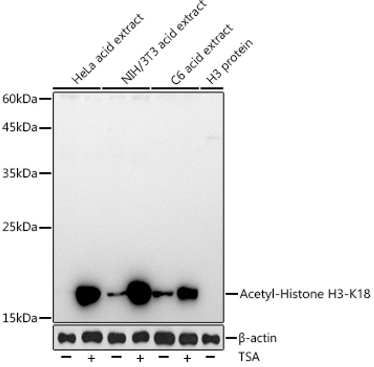
Western blot analysis of extracts of various cell lines, using Acetyl-Histone H3-K18 rabbit monoclonal antibody (A20680) at 1:1000 dilution. HeLa cells and NIH/3T3 cells and C6 cells were treated by TSA (1 uM) at 37 °C for 18 hours. Secondary antibody: HRP Goat Anti-rabbit IgG (H+L) (STJS000856) at 1:10000 dilution. Lysates/proteins: 25 Mu g per lane. Blocking buffer: 3% non-fat dry milk in TBST. Detection: ECL Basic Kit. Exposure time: 180s.
-
Dot-blot analysis of all sorts of peptides using Acetyl-Histone H3-K18 rabbit monoclonal antibody (STJ11103433) at 1:100000 dilution.
-
Immunohistochemistry analysis of Acetyl-Histone H3-K18 in paraffin-embedded human cervix cancer tissue using Acetyl-Histone H3-K18 rabbit monoclonal antibody (STJ11103433) at a dilution of 1:200 (40x lens). High pressure antigen retrieval was performed with 0. 01 M citrate buffer (pH 6. 0) prior to immunohistochemistry staining.
-
Immunohistochemistry analysis of Acetyl-Histone H3-K18 in paraffin-embedded human colon carcinoma tissue using Acetyl-Histone H3-K18 rabbit monoclonal antibody (STJ11103433) at a dilution of 1:200 (40x lens). High pressure antigen retrieval was performed with 0. 01 M citrate buffer (pH 6. 0) prior to immunohistochemistry staining.
-
Immunohistochemistry analysis of Acetyl-Histone H3-K18 in paraffin-embedded human colon tissue using Acetyl-Histone H3-K18 rabbit monoclonal antibody (STJ11103433) at a dilution of 1:200 (40x lens). High pressure antigen retrieval was performed with 0. 01 M citrate buffer (pH 6. 0) prior to immunohistochemistry staining.
-
Immunohistochemistry analysis of Acetyl-Histone H3-K18 in paraffin-embedded mouse brain tissue using Acetyl-Histone H3-K18 rabbit monoclonal antibody (STJ11103433) at a dilution of 1:200 (40x lens). High pressure antigen retrieval was performed with 0. 01 M citrate buffer (pH 6. 0) prior to immunohistochemistry staining.
-
Immunohistochemistry analysis of Acetyl-Histone H3-K18 in paraffin-embedded mouse intestin tissue using Acetyl-Histone H3-K18 rabbit monoclonal antibody (STJ11103433) at a dilution of 1:200 (40x lens). High pressure antigen retrieval was performed with 0. 01 M citrate buffer (pH 6. 0) prior to immunohistochemistry staining.
-
Immunohistochemistry analysis of Acetyl-Histone H3-K18 in paraffin-embedded mouse kidney tissue using Acetyl-Histone H3-K18 rabbit monoclonal antibody (STJ11103433) at a dilution of 1:200 (40x lens). High pressure antigen retrieval was performed with 0. 01 M citrate buffer (pH 6. 0) prior to immunohistochemistry staining.
-
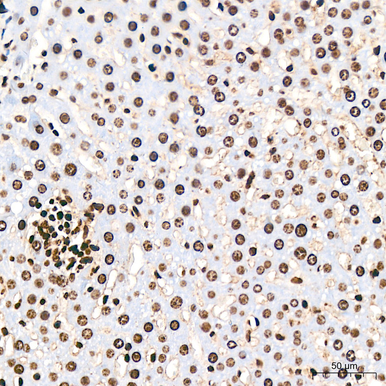
Immunohistochemistry analysis of Acetyl-Histone H3-K18 in paraffin-embedded mouse liver tissue using Acetyl-Histone H3-K18 rabbit monoclonal antibody (STJ11103433) at a dilution of 1:200 (40x lens). High pressure antigen retrieval was performed with 0. 01 M citrate buffer (pH 6. 0) prior to immunohistochemistry staining.
-
Immunohistochemistry analysis of Acetyl-Histone H3-K18 in paraffin-embedded mouse lung tissue using Acetyl-Histone H3-K18 rabbit monoclonal antibody (STJ11103433) at a dilution of 1:200 (40x lens). High pressure antigen retrieval was performed with 0. 01 M citrate buffer (pH 6. 0) prior to immunohistochemistry staining.
-
Immunohistochemistry analysis of Acetyl-Histone H3-K18 in paraffin-embedded mouse spleen tissue using Acetyl-Histone H3-K18 rabbit monoclonal antibody (STJ11103433) at a dilution of 1:200 (40x lens). High pressure antigen retrieval was performed with 0. 01 M citrate buffer (pH 6. 0) prior to immunohistochemistry staining.
-
Immunohistochemistry analysis of Acetyl-Histone H3-K18 in paraffin-embedded rat brain tissue using Acetyl-Histone H3-K18 rabbit monoclonal antibody (STJ11103433) at a dilution of 1:200 (40x lens). High pressure antigen retrieval was performed with 0. 01 M citrate buffer (pH 6. 0) prior to immunohistochemistry staining.
-
Immunohistochemistry analysis of Acetyl-Histone H3-K18 in paraffin-embedded rat colon tissue using Acetyl-Histone H3-K18 rabbit monoclonal antibody (STJ11103433) at a dilution of 1:200 (40x lens). High pressure antigen retrieval was performed with 0. 01 M citrate buffer (pH 6. 0) prior to immunohistochemistry staining.
-
Immunohistochemistry analysis of Acetyl-Histone H3-K18 in paraffin-embedded rat liver tissue using Acetyl-Histone H3-K18 rabbit monoclonal antibody (STJ11103433) at a dilution of 1:200 (40x lens). High pressure antigen retrieval was performed with 0. 01 M citrate buffer (pH 6. 0) prior to immunohistochemistry staining.
-
Immunohistochemistry analysis of Acetyl-Histone H3-K18 in paraffin-embedded rat lung tissue using Acetyl-Histone H3-K18 rabbit monoclonal antibody (STJ11103433) at a dilution of 1:200 (40x lens). High pressure antigen retrieval was performed with 0. 01 M citrate buffer (pH 6. 0) prior to immunohistochemistry staining.
-
Chromatin immunoprecipitation analysis of extracts of HeLa cells, using Acetyl-Histone H3-K18 rabbit monoclonal antibody (STJ11103433) and rabbit IgG. The amount of immunoprecipitated DNA was checked by quantitative PCR. Histogram was constructed by the ratios of the immunoprecipitated DNA to the input.


