| Host: |
Mouse |
| Applications: |
WB/IHC/IF/IP |
| Reactivity: |
Human |
| Note: |
STRICTLY FOR FURTHER SCIENTIFIC RESEARCH USE ONLY (RUO). MUST NOT TO BE USED IN DIAGNOSTIC OR THERAPEUTIC APPLICATIONS. |
| Short Description: |
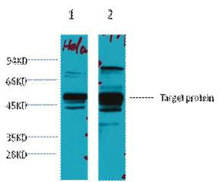
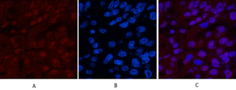
Mouse monoclonal antibody anti-DNA repair protein XRCC4 is suitable for use in Western Blot, Immunohistochemistry, Immunofluorescence and Immunoprecipitation research applications. |
| Clonality: |
Monoclonal |
| Clone ID: |
5C10 |
| Conjugation: |
Unconjugated |
| Isotype: |
IgG1 |
| Formulation: |
Liquid in PBS pH7.4, 0.5% BSA, 0.02% Sodium Azide and 50% Glycerol. |
| Purification: |
The antibody was affinity-purified from mouse ascites by affinity-chromatography using specific immunogen. |
| Dilution Range: |
WB 1:2000IP 1:200IF 1:200IHC 1:50-300 |
| Storage Instruction: |
Store at-20°C for up to 1 year from the date of receipt, and avoid repeat freeze-thaw cycles. |
| Gene Symbol: |
XRCC4 |
| Gene ID: |
7518 |
| Uniprot ID: |
XRCC4_HUMAN |
| Specificity: |
The antibody detects endogenous XRCC4 proteins. |
| Immunogen: |
Synthetic Peptide of XRCC4 |
| Post Translational Modifications | Phosphorylated by PRKDC at the C-terminus in response to DNA damage.Ser-260 and Ser-320 constitute the main phosphorylation sites. Phosphorylations by PRKDC at the C-terminus of XRCC4 and NHEJ1/XLF are highly redundant and regulate ability of the XRCC4-NHEJ1/XLF subcomplex to bridge DNA. Phosphorylation by PRKDC does not prevent interaction with NHEJ1/XLF but disrupts ability to bridge DNA and promotes detachment from DNA. Phosphorylation at Ser-327 and Ser-328 by PRKDC promotes recognition by the SCF(FBXW7) complex and subsequent ubiquitination via 'Lys-63'-linked ubiquitin. Phosphorylation at Thr-233 by CK2 promotes interaction with PNKP.regulating PNKP activity and localization to DNA damage sites. Phosphorylation by CK2 promotes interaction with APTX. Ubiquitinated at Lys-296 by the SCF(FBXW7) complex via 'Lys-63'-linked ubiquitination, thereby promoting double-strand break repair: the SCF(FBXW7) complex specifically recognizes XRCC4 when phosphorylated at Ser-327 and Ser-328 by PRKDC, and 'Lys-63'-linked ubiquitination facilitates DNA non-homologous end joining (NHEJ) by enhancing association with XRCC5/Ku80 and XRCC6/Ku70. Monoubiquitinated. DNA repair protein XRCC4: Undergoes proteolytic processing by caspase-3 (CASP3) (Probable). This generates the protein XRCC4, C-terminus (XRCC4/C), which translocates to the cytoplasm and activates phospholipid scramblase activity of XKR4, thereby promoting phosphatidylserine exposure on apoptotic cell surface. |
| Function | DNA repair protein XRCC4: DNA non-homologous end joining (NHEJ) core factor, required for double-strand break repair and V(D)J recombination. Acts as a scaffold protein that regulates recruitment of other proteins to DNA double-strand breaks (DSBs). Associates with NHEJ1/XLF to form alternating helical filaments that bridge DNA and act like a bandage, holding together the broken DNA until it is repaired. The XRCC4-NHEJ1/XLF subcomplex binds to the DNA fragments of a DSB in a highly diffusive manner and robustly bridges two independent DNA molecules, holding the broken DNA fragments in close proximity to one other. The mobility of the bridges ensures that the ends remain accessible for further processing by other repair factors. Plays a key role in the NHEJ ligation step of the broken DNA during DSB repair via direct interaction with DNA ligase IV (LIG4): the LIG4-XRCC4 subcomplex reseals the DNA breaks after the gap filling is completed. XRCC4 stabilizes LIG4, regulates its subcellular localization and enhances LIG4's joining activity. Binding of the LIG4-XRCC4 subcomplex to DNA ends is dependent on the assembly of the DNA-dependent protein kinase complex DNA-PK to these DNA ends. Promotes displacement of PNKP from processed strand break termini. Protein XRCC4, C-terminus: Acts as an activator of the phospholipid scramblase activity of XKR4. This form, which is generated upon caspase-3 (CASP3) cleavage, translocates into the cytoplasm and interacts with XKR4, thereby promoting phosphatidylserine scramblase activity of XKR4 and leading to phosphatidylserine exposure on apoptotic cell surface. |
| Protein Name | Dna Repair Protein Xrcc4Hxrcc4X-Ray Repair Cross-Complementing Protein 4 Cleaved Into - Protein Xrcc4 - C-TerminusXrcc4/C |
| Database Links | Reactome: R-HSA-164843Reactome: R-HSA-3108214Reactome: R-HSA-5693571 |
| Cellular Localisation | NucleusChromosomeLocalizes To Site Of Double-Strand BreaksProtein Xrcc4C-Terminus: CytoplasmTranslocates From The Nucleus To The Cytoplasm Following Cleavage By Caspase-3 (Casp3) |
| Alternative Antibody Names | Anti-Dna Repair Protein Xrcc4 antibodyAnti-Hxrcc4 antibodyAnti-X-Ray Repair Cross-Complementing Protein 4 Cleaved Into - Protein Xrcc4 - C-Terminus antibodyAnti-Xrcc4/C antibodyAnti-XRCC4 antibody |
Information sourced from Uniprot.org
12 months for antibodies. 6 months for ELISA Kits. Please see website T&Cs for further guidance


![Western blot analysis of lysates from wild type (WT) and XRCC4 knockout (KO) HeLa cells, using [KO Validated] XRCC4 Rabbit polyclonal antibody (STJ11100019) at 1:500 dilution. Secondary antibody: HRP Goat Anti-Rabbit IgG (H+L) (STJS000856) at 1:10000 dilution. Lysates/proteins: 25 Mu g per lane. Blocking buffer: 3% nonfat dry milk in TBST. Detection: ECL Basic Kit. Exposure time: 30s. Western blot analysis of lysates from wild type (WT) and XRCC4 knockout (KO) HeLa cells, using [KO Validated] XRCC4 Rabbit polyclonal antibody (STJ11100019) at 1:500 dilution. Secondary antibody: HRP Goat Anti-Rabbit IgG (H+L) (STJS000856) at 1:10000 dilution. Lysates/proteins: 25 Mu g per lane. Blocking buffer: 3% nonfat dry milk in TBST. Detection: ECL Basic Kit. Exposure time: 30s.](https://cdn11.bigcommerce.com/s-zso2xnchw9/images/stencil/300x300/products/89092/357551/STJ11100019_1__20826.1713121671.jpg?c=1)


