| Host: |
Rabbit |
| Applications: |
WB/IF |
| Reactivity: |
Human/Mouse/Rat |
| Note: |
STRICTLY FOR FURTHER SCIENTIFIC RESEARCH USE ONLY (RUO). MUST NOT TO BE USED IN DIAGNOSTIC OR THERAPEUTIC APPLICATIONS. |
| Short Description: |
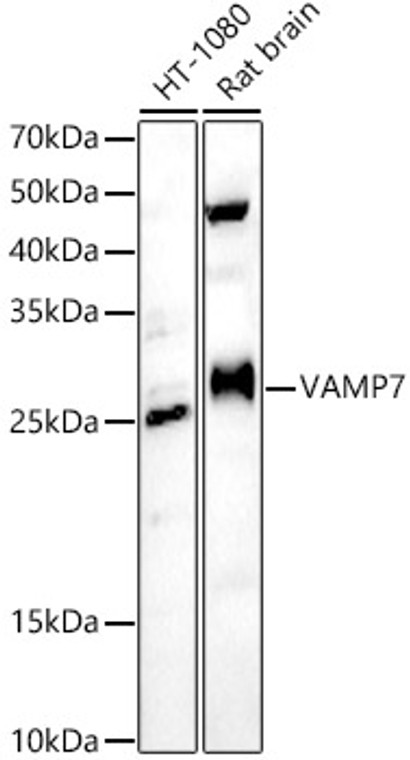
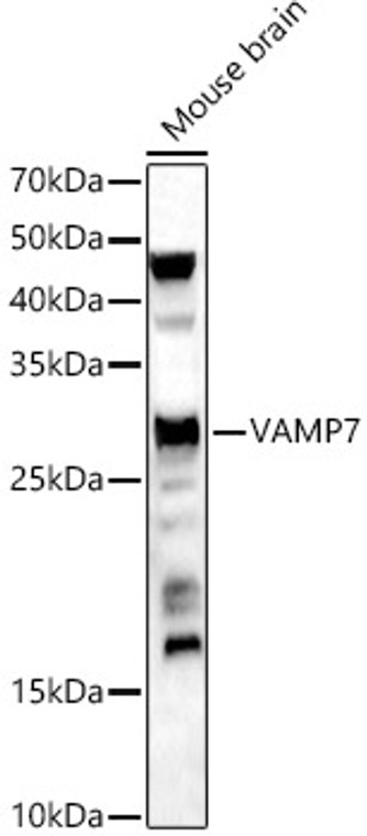
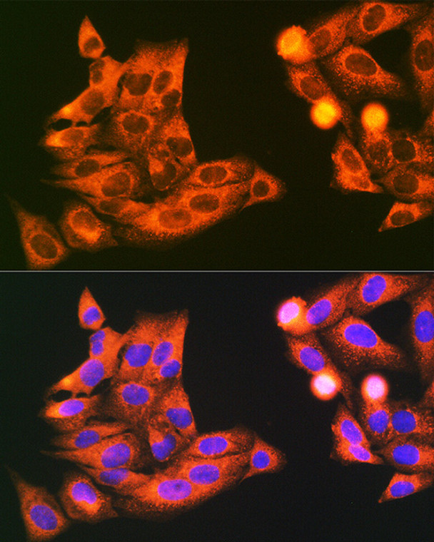
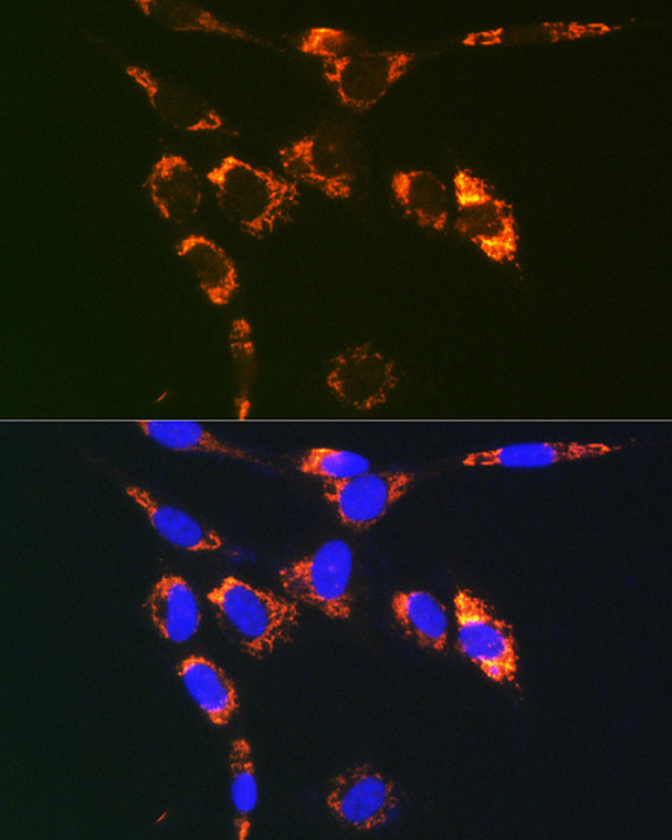
Rabbit polyclonal antibody anti-VAMP7 (11-70) is suitable for use in Western Blot and Immunofluorescence research applications. |
| Clonality: |
Polyclonal |
| Conjugation: |
Unconjugated |
| Isotype: |
IgG |
| Formulation: |
PBS with 0.01% Thimerosal, 50% Glycerol, pH7.3. |
| Purification: |
Affinity purification |
| Dilution Range: |
WB 1:100-1:500IF/ICC 1:50-1:200 |
| Storage Instruction: |
Store at-20°C for up to 1 year from the date of receipt, and avoid repeat freeze-thaw cycles. |
| Gene Symbol: |
VAMP7 |
| Gene ID: |
6845 |
| Uniprot ID: |
VAMP7_HUMAN |
| Immunogen Region: |
11-70 |
| Immunogen: |
Recombinant fusion protein containing a sequence corresponding to amino acids 11-70 of human VAMP7 (P51809). |
| Immunogen Sequence: |
GTTILAKHAWCGGNFLEVTE QILAKIPSENNKLTYSHGNY LFHYICQDRIVYLCITDDDF |
| Tissue Specificity | Detected in all tissues tested. |
| Function | Involved in the targeting and/or fusion of transport vesicles to their target membrane during transport of proteins from the early endosome to the lysosome. Required for heterotypic fusion of late endosomes with lysosomes and homotypic lysosomal fusion. Required for calcium regulated lysosomal exocytosis. Involved in the export of chylomicrons from the endoplasmic reticulum to the cis Golgi. Required for exocytosis of mediators during eosinophil and neutrophil degranulation, and target cell killing by natural killer cells. Required for focal exocytosis of late endocytic vesicles during phagosome formation. |
| Protein Name | Vesicle-Associated Membrane Protein 7Vamp-7Synaptobrevin-Like Protein 1Tetanus-Insensitive VampTi-Vamp |
| Database Links | Reactome: R-HSA-199992Reactome: R-HSA-432720Reactome: R-HSA-432722Reactome: R-HSA-8856825Reactome: R-HSA-8856828Reactome: R-HSA-9020591 |
| Cellular Localisation | Cytoplasmic VesicleSecretory Vesicle MembraneSingle-Pass Type Iv Membrane ProteinGolgi ApparatusTrans-Golgi Network MembraneLate Endosome MembraneLysosome MembraneEndoplasmic Reticulum MembranePhagosome MembraneSynapseSynaptosomeIn Immature Neurons Expression Is Localized In Vesicular Structures In Axons And Dendrites While In Mature Neurons It Is Localized To The Somatodendritic RegionColocalizes With Lamp1 In Kidney CellsLocalization To The Endoplasmic Reticulum Membrane Was Observed In The Intestine But Not In Liver Or Kidney |
| Alternative Antibody Names | Anti-Vesicle-Associated Membrane Protein 7 antibodyAnti-Vamp-7 antibodyAnti-Synaptobrevin-Like Protein 1 antibodyAnti-Tetanus-Insensitive Vamp antibodyAnti-Ti-Vamp antibodyAnti-VAMP7 antibodyAnti-SYBL1 antibody |
Information sourced from Uniprot.org
12 months for antibodies. 6 months for ELISA Kits. Please see website T&Cs for further guidance


