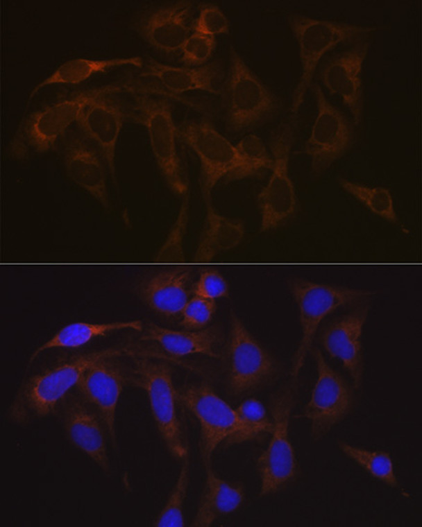
| Host: |
Rabbit |
| Applications: |
WB/IHC/IF |
| Reactivity: |
Human/Mouse/Rat |
| Note: |
STRICTLY FOR FURTHER SCIENTIFIC RESEARCH USE ONLY (RUO). MUST NOT TO BE USED IN DIAGNOSTIC OR THERAPEUTIC APPLICATIONS. |
| Short Description: |
Rabbit monoclonal antibody anti-VAMP1 (1-118) is suitable for use in Western Blot, Immunohistochemistry and Immunofluorescence research applications. |
| Clonality: |
Monoclonal |
| Clone ID: |
S2MR |
| Conjugation: |
Unconjugated |
| Isotype: |
IgG |
| Formulation: |
PBS with 0.02% Sodium Azide, 0.05% BSA, 50% Glycerol, pH7.3. |
| Purification: |
Affinity purification |
| Dilution Range: |
WB 1:500-1:2000IHC-P 1:50-1:200IF/ICC 1:50-1:200 |
| Storage Instruction: |
Store at-20°C for up to 1 year from the date of receipt, and avoid repeat freeze-thaw cycles. |
| Gene Symbol: |
VAMP1 |
| Gene ID: |
6843 |
| Uniprot ID: |
VAMP1_HUMAN |
| Immunogen Region: |
1-118 |
| Immunogen: |
Recombinant fusion protein containing a sequence corresponding to amino acids 1-118 of human VAMP1 (P23763). |
| Immunogen Sequence: |
MSAPAQPPAEGTEGTAPGGG PPGPPPNMTSNRRLQQTQAQ VEEVVDIIRVNVDKVLERDQ KLSELDDRADALQAGASQFE SSAAKLKRKYWWKNCKMMIM LGAICAIIVVVIVIYFFT |
| Tissue Specificity | Nervous system, skeletal muscle and adipose tissue. |
| Post Translational Modifications | (Microbial infection) Targeted and hydrolyzed by C.botulinum neurotoxin type B (BoNT/B, botB) which probably hydrolyzes the 78-Gln-|-Phe-79 bond and inhibits neurotransmitter release. (Microbial infection) Targeted and hydrolyzed by C.botulinum neurotoxin type D (BoNT/D, botD) which probably hydrolyzes the 61-Arg-|-Leu-62 bond and inhibits neurotransmitter release. BoNT/D has low catalytic activity on this protein due to its sequence. Note that humans are not known to be infected by C.botulinum type D. (Microbial infection) Targeted and hydrolyzed by C.botulinum neurotoxin type F (BoNT/F, botF) which probably hydrolyzes the 60-Gln-|-Lys-61 bond and inhibits neurotransmitter release. (Microbial infection) Targeted and hydrolyzed by C.botulinum neurotoxin type X (BoNT/X) which probably hydrolyzes the 68-Arg-|-Ala-69 bond and inhibits neurotransmitter release. It remains unknown whether BoNT/X is ever produced, or what organisms it targets. |
| Function | Involved in the targeting and/or fusion of transport vesicles to their target membrane. |
| Protein Name | Vesicle-Associated Membrane Protein 1Vamp-1Synaptobrevin-1 |
| Database Links | Reactome: R-HSA-5250955Reactome: R-HSA-5250981Reactome: R-HSA-5250989 |
| Cellular Localisation | Isoform 1: Cytoplasmic VesicleSecretory VesicleSynaptic Vesicle MembraneSingle-Pass Type Iv Membrane ProteinSynapseSynaptosomeIsoform 2: Cytoplasmic Vesicle MembraneIsoform 3: Mitochondrion Outer Membrane |
| Alternative Antibody Names | Anti-Vesicle-Associated Membrane Protein 1 antibodyAnti-Vamp-1 antibodyAnti-Synaptobrevin-1 antibodyAnti-VAMP1 antibodyAnti-SYB1 antibody |
Information sourced from Uniprot.org
12 months for antibodies. 6 months for ELISA Kits. Please see website T&Cs for further guidance


