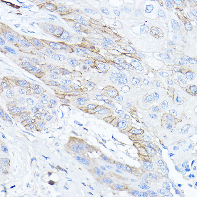
| Host: |
Rabbit |
| Applications: |
WB/IHC/IF |
| Reactivity: |
Human/Mouse/Rat |
| Note: |
STRICTLY FOR FURTHER SCIENTIFIC RESEARCH USE ONLY (RUO). MUST NOT TO BE USED IN DIAGNOSTIC OR THERAPEUTIC APPLICATIONS. |
| Short Description: |
Rabbit polyclonal antibody anti-TMPRSS2 (1-100) is suitable for use in Western Blot, Immunohistochemistry and Immunofluorescence research applications. |
| Clonality: |
Polyclonal |
| Conjugation: |
Unconjugated |
| Isotype: |
IgG |
| Formulation: |
PBS with 0.05% Proclin300, 50% Glycerol, pH7.3. |
| Purification: |
Affinity purification |
| Dilution Range: |
WB 1:500-1:1000IHC-P 1:50-1:200IF/ICC 1:50-1:200 |
| Storage Instruction: |
Store at-20°C for up to 1 year from the date of receipt, and avoid repeat freeze-thaw cycles. |
| Gene Symbol: |
TMPRSS2 |
| Gene ID: |
7113 |
| Uniprot ID: |
TMPS2_HUMAN |
| Immunogen Region: |
1-100 |
| Immunogen: |
A synthetic peptide corresponding to a sequence within amino acids 1-100 of human TMPRSS2 (NP_005647.3). |
| Immunogen Sequence: |
MALNSGSPPAIGPYYENHGY QPENPYPAQPTVVPTVYEVH PAQYYPSPVPQYAPRVLTQA SNPVVCTQPKSPSGTVCTSK TKKALCITLTLGTFLVGAAL |
| Tissue Specificity | Expressed in several tissues that comprise large populations of epithelial cells with the highest level of transcripts measured in the prostate gland. Expressed in type II pneumocytes in the lung (at protein level). Expressed strongly in small intestine. Also expressed in colon, stomach and salivary gland. Coexpressed with ACE2 within lung type II pneumocytes, ileal absorptive enterocytes, intestinal epithelial cells, cornea, gallbladder and nasal goblet secretory cells (Ref.21). |
| Post Translational Modifications | Proteolytically processed.by an autocatalytic mechanism. |
| Function | Plasma membrane-anchored serine protease that cleaves at arginine residues. Participates in proteolytic cascades of relevance for the normal physiologic function of the prostate. Androgen-induced TMPRSS2 activates several substrates that include pro-hepatocyte growth factor/HGF, the protease activated receptor-2/F2RL1 or matriptase/ST14 leading to extracellular matrix disruption and metastasis of prostate cancer cells. In addition, activates trigeminal neurons and contribute to both spontaneous pain and mechanical allodynia. (Microbial infection) Facilitates human coronaviruses SARS-CoV and SARS-CoV-2 infections via two independent mechanisms, proteolytic cleavage of ACE2 receptor which promotes viral uptake, and cleavage of coronavirus spike glycoproteins which activates the glycoprotein for host cell entry. The cleavage of SARS-COV2 spike glycoprotein occurs between the S2 and S2' site. Upon SARS-CoV-2 infection, increases syncytia formation by accelerating the fusion process. Proteolytically cleaves and activates the spike glycoproteins of human coronavirus 229E (HCoV-229E) and human coronavirus EMC (HCoV-EMC) and the fusion glycoproteins F0 of Sendai virus (SeV), human metapneumovirus (HMPV), human parainfluenza 1, 2, 3, 4a and 4b viruses (HPIV). Essential for spread and pathogenesis of influenza A virus (strains H1N1, H3N2 and H7N9).involved in proteolytic cleavage and activation of hemagglutinin (HA) protein which is essential for viral infectivity. |
| Protein Name | Transmembrane Protease Serine 2Serine Protease 10 Cleaved Into - Transmembrane Protease Serine 2 Non-Catalytic Chain - Transmembrane Protease Serine 2 Catalytic Chain |
| Database Links | Reactome: R-HSA-9678110Reactome: R-HSA-9694614Reactome: R-HSA-9733458 |
| Cellular Localisation | Cell MembraneSingle-Pass Type Ii Membrane ProteinTransmembrane Protease Serine 2 Catalytic Chain: SecretedActivated By Cleavage And Secreted |
| Alternative Antibody Names | Anti-Transmembrane Protease Serine 2 antibodyAnti-Serine Protease 10 Cleaved Into - Transmembrane Protease Serine 2 Non-Catalytic Chain - Transmembrane Protease Serine 2 Catalytic Chain antibodyAnti-TMPRSS2 antibodyAnti-PRSS10 antibody |
Information sourced from Uniprot.org
12 months for antibodies. 6 months for ELISA Kits. Please see website T&Cs for further guidance


