| Host: |
Rabbit |
| Applications: |
WB/IHC |
| Reactivity: |
Human/Mouse/Rat |
| Note: |
STRICTLY FOR FURTHER SCIENTIFIC RESEARCH USE ONLY (RUO). MUST NOT TO BE USED IN DIAGNOSTIC OR THERAPEUTIC APPLICATIONS. |
| Short Description: |
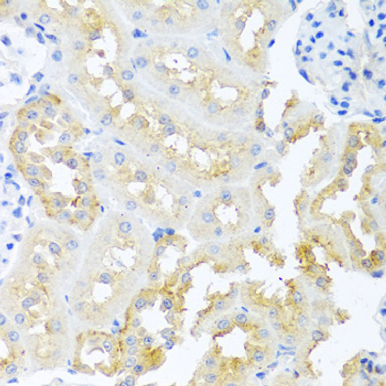
Rabbit polyclonal antibody anti-TDGF1 (31-150) is suitable for use in Western Blot and Immunohistochemistry research applications. |
| Clonality: |
Polyclonal |
| Conjugation: |
Unconjugated |
| Isotype: |
IgG |
| Formulation: |
PBS with 0.05% Proclin300, 50% Glycerol, pH7.3. |
| Purification: |
Affinity purification |
| Dilution Range: |
WB 1:500-1:1000IHC-P 1:50-1:200 |
| Storage Instruction: |
Store at-20°C for up to 1 year from the date of receipt, and avoid repeat freeze-thaw cycles. |
| Gene Symbol: |
CRIPTO |
| Gene ID: |
6997 |
| Uniprot ID: |
TDGF1_HUMAN |
| Immunogen Region: |
31-150 |
| Immunogen: |
Recombinant fusion protein containing a sequence corresponding to amino acids 31-150 of human TDGF1 (NP_001167607.1). |
| Immunogen Sequence: |
RDDSIWPQEEPAIRPRSSQR VPPMGIQHSKELNRTCCLNG GTCMLGSFCACPPSFYGRNC EHDVRKENCGSVPHDTWLPK KCSLCKCWHGQLRCFPQAFL PGCDGLVMDEHLVASRTPEL |
| Tissue Specificity | Preferentially expressed in gastric and colorectal carcinomas than in their normal counterparts. Expressed in breast and lung. |
| Post Translational Modifications | The GPI-anchor is attached to the protein in the endoplasmic reticulum and serves to target the protein to the cell surface. There, it is processed by GPI processing phospholipase A2 (TMEM8A), removing an acyl-chain at the sn-2 position of GPI and releasing CRIPTO as a lysophosphatidylinositol-bearing form, which is further cleaved by phospholipase D (GPLD1) into a soluble form. |
| Function | GPI-anchored cell membrane protein involved in Nodal signaling. Cell-associated CRIPTO acts as a Nodal coreceptor in cis. Shedding of CRIPTO by TMEM8A modulates Nodal signaling by allowing soluble CRIPTO to act as a Nodal coreceptor on other cells. Could play a role in the determination of the epiblastic cells that subsequently give rise to the mesoderm. |
| Protein Name | Protein CriptoCripto - Egf-Cfc Family MemberCripto-1 Growth FactorCrgfEpidermal Growth Factor-Like Cripto Protein Cr1Teratocarcinoma-Derived Growth Factor 1 |
| Database Links | Reactome: R-HSA-1181150Reactome: R-HSA-1433617Reactome: R-HSA-2892247 |
| Cellular Localisation | Cell MembraneLipid-AnchorGpi-AnchorSecretedReleased From The Cell Membrane By Gpi Cleavage |
| Alternative Antibody Names | Anti-Protein Cripto antibodyAnti-Cripto - Egf-Cfc Family Member antibodyAnti-Cripto-1 Growth Factor antibodyAnti-Crgf antibodyAnti-Epidermal Growth Factor-Like Cripto Protein Cr1 antibodyAnti-Teratocarcinoma-Derived Growth Factor 1 antibodyAnti-CRIPTO antibodyAnti-CRIPTO-1 antibodyAnti-TDGF1 antibody |
Information sourced from Uniprot.org
12 months for antibodies. 6 months for ELISA Kits. Please see website T&Cs for further guidance


