| Host: |
Rabbit |
| Applications: |
WB/IHC/IF |
| Reactivity: |
Human/Mouse/Rat |
| Note: |
STRICTLY FOR FURTHER SCIENTIFIC RESEARCH USE ONLY (RUO). MUST NOT TO BE USED IN DIAGNOSTIC OR THERAPEUTIC APPLICATIONS. |
| Short Description: |
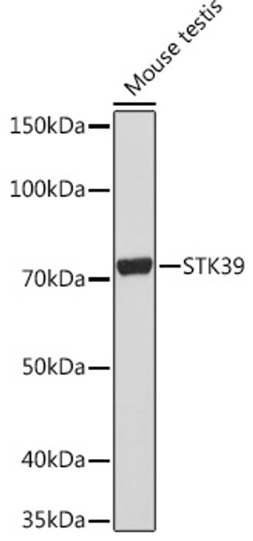
Rabbit monoclonal antibody anti-STK39 (401-545) is suitable for use in Western Blot, Immunohistochemistry and Immunofluorescence research applications. |
| Clonality: |
Monoclonal |
| Clone ID: |
S2MR |
| Conjugation: |
Unconjugated |
| Isotype: |
IgG |
| Formulation: |
PBS with 0.02% Sodium Azide, 0.05% BSA, 50% Glycerol, pH7.3. |
| Purification: |
Affinity purification |
| Dilution Range: |
WB 1:500-1:2000IHC-P 1:50-1:200IF/ICC 1:50-1:200 |
| Storage Instruction: |
Store at-20°C for up to 1 year from the date of receipt, and avoid repeat freeze-thaw cycles. |
| Gene Symbol: |
STK39 |
| Gene ID: |
27347 |
| Uniprot ID: |
STK39_HUMAN |
| Immunogen Region: |
401-545 |
| Immunogen: |
Recombinant fusion protein containing a sequence corresponding to amino acids 401-545 of human STK39 (Q9UEW8). |
| Immunogen Sequence: |
SQEKSRRVKEENPEIAVSAS TIPEQIQSLSVHDSQGPPNA NEDYREASSCAVNLVLRLRN SRKELNDIRFEFTPGRDTAD GVSQELFSAGLVDGHDVVIV AANLQKIVDDPKALKTLTFK LASGCDGSEIPDEVKLIGFA QLSVS |
| Tissue Specificity | Predominantly expressed in brain and pancreas followed by heart, lung, kidney, skeletal muscle, liver, placenta and testis. |
| Post Translational Modifications | Phosphorylation at Thr-231 by WNK kinases (WNK1, WNK2, WNK3 or WNK4) is required for activation. Autophosphorylation at Thr-231 positively regulates its activity. Phosphorylated at Ser-309 by PRKCQ. |
| Function | Effector serine/threonine-protein kinase component of the WNK-SPAK/OSR1 kinase cascade, which is involved in various processes, such as ion transport, response to hypertonic stress and blood pressure. Specifically recognizes and binds proteins with a RFXV motif. Acts downstream of WNK kinases (WNK1, WNK2, WNK3 or WNK4): following activation by WNK kinases, catalyzes phosphorylation of ion cotransporters, such as SLC12A1/NKCC2, SLC12A2/NKCC1, SLC12A3/NCC, SLC12A5/KCC2 or SLC12A6/KCC3, regulating their activity. Mediates regulatory volume increase in response to hyperosmotic stress by catalyzing phosphorylation of ion cotransporters SLC12A1/NKCC2, SLC12A2/NKCC1 and SLC12A6/KCC3 downstream of WNK1 and WNK3 kinases. Phosphorylation of Na-K-Cl cotransporters SLC12A2/NKCC1 and SLC12A2/NKCC1 promote their activation and ion influx.simultaneously, phosphorylation of K-Cl cotransporters SLC12A5/KCC2 and SLC12A6/KCC3 inhibit their activity, blocking ion efflux. Acts as a regulator of NaCl reabsorption in the distal nephron by mediating phosphorylation and activation of the thiazide-sensitive Na-Cl cotransporter SLC12A3/NCC in distal convoluted tubule cells of kidney downstream of WNK4. Mediates the inhibition of SLC4A4, SLC26A6 as well as CFTR activities. Phosphorylates RELT. |
| Protein Name | Ste20/Sps1-Related Proline-Alanine-Rich Protein KinaseSte-20-Related KinaseDchtSerine/Threonine-Protein Kinase 39 |
| Cellular Localisation | CytoplasmNucleusNucleus When Caspase-Cleaved |
| Alternative Antibody Names | Anti-Ste20/Sps1-Related Proline-Alanine-Rich Protein Kinase antibodyAnti-Ste-20-Related Kinase antibodyAnti-Dcht antibodyAnti-Serine/Threonine-Protein Kinase 39 antibodyAnti-STK39 antibodyAnti-PASK antibodyAnti-SPAK antibody |
Information sourced from Uniprot.org
12 months for antibodies. 6 months for ELISA Kits. Please see website T&Cs for further guidance


