| Host: |
Rabbit |
| Applications: |
WB/IHC |
| Reactivity: |
Human/Mouse/Rat |
| Note: |
STRICTLY FOR FURTHER SCIENTIFIC RESEARCH USE ONLY (RUO). MUST NOT TO BE USED IN DIAGNOSTIC OR THERAPEUTIC APPLICATIONS. |
| Short Description: |
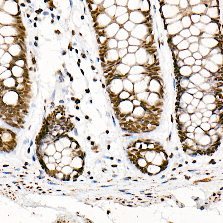
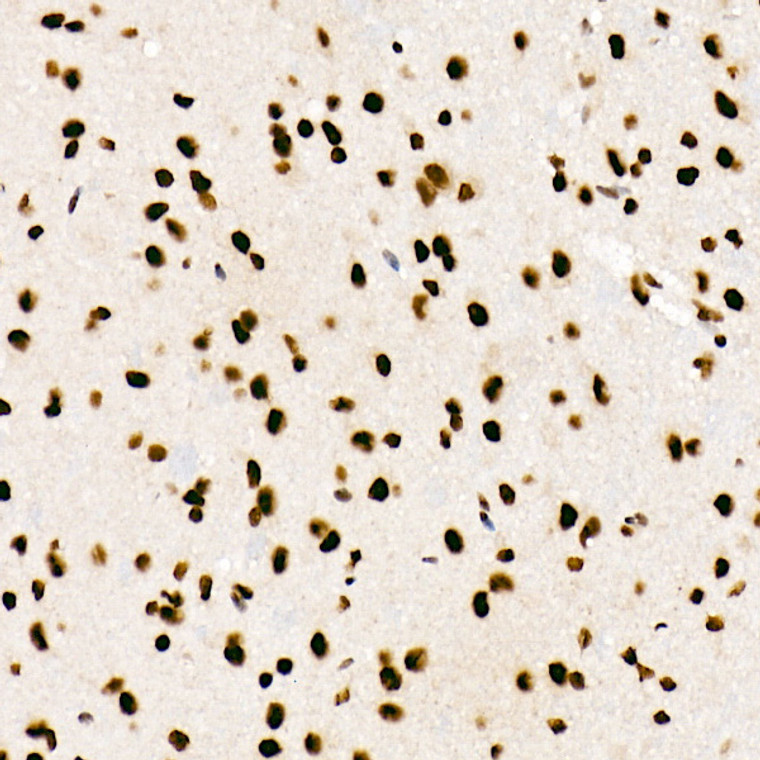
Rabbit monoclonal antibody anti-SF2/ASF (100-200) is suitable for use in Western Blot and Immunohistochemistry research applications. |
| Clonality: |
Monoclonal |
| Clone ID: |
S5MR |
| Conjugation: |
Unconjugated |
| Isotype: |
IgG |
| Formulation: |
PBS with 0.05% Proclin300, 0.05% BSA, 50% Glycerol, pH7.3. |
| Purification: |
Affinity purification |
| Dilution Range: |
WB 1:500-1:1000IHC-P 1:50-1:200 |
| Storage Instruction: |
Store at-20°C for up to 1 year from the date of receipt, and avoid repeat freeze-thaw cycles. |
| Gene Symbol: |
SRSF1 |
| Gene ID: |
6426 |
| Uniprot ID: |
SRSF1_HUMAN |
| Immunogen Region: |
100-200 |
| Immunogen: |
A synthetic peptide corresponding to a sequence within amino acids 100-200 of human SRSF1/SF2/ASF (NP_008855.1). |
| Immunogen Sequence: |
GGGGGGGAPRGRYGPPSRRS ENRVVVSGLPPSGSWQDLKD HMREAGDVCYADVYRDGTGV VEFVRKEDMTYAVRKLDNTK FRSHEGETAYIRVKVDGPRS P |
| Post Translational Modifications | Phosphorylated by CLK1, CLK2, CLK3 and CLK4. Phosphorylated by SRPK1 at multiple serines in its RS domain via a directional (C-terminal to N-terminal) and a dual-track mechanism incorporating both processive phosphorylation (in which the kinase stays attached to the substrate after each round of phosphorylation) and distributive phosphorylation steps (in which the kinase and substrate dissociate after each phosphorylation event). The RS domain of SRSF1 binds to a docking groove in the large lobe of the kinase domain of SRPK1 and this induces certain structural changes in SRPK1 and/or RRM 2 domain of SRSF1, allowing RRM 2 to bind the kinase and initiate phosphorylation. The cycles continue for several phosphorylation steps in a processive manner (steps 1-8) until the last few phosphorylation steps (approximately steps 9-12). During that time, a mechanical stress induces the unfolding of the beta-4 motif in RRM 2, which then docks at the docking groove of SRPK1. This also signals RRM 2 to begin to dissociate, which facilitates SRSF1 dissociation after phosphorylation is completed. Asymmetrically dimethylated at arginines, probably by PRMT1, methylation promotes localization to nuclear speckles. |
| Function | Plays a role in preventing exon skipping, ensuring the accuracy of splicing and regulating alternative splicing. Interacts with other spliceosomal components, via the RS domains, to form a bridge between the 5'- and 3'-splice site binding components, U1 snRNP and U2AF. Can stimulate binding of U1 snRNP to a 5'-splice site-containing pre-mRNA. Binds to purine-rich RNA sequences, either the octamer, 5'-RGAAGAAC-3' (r=A or G) or the decamers, AGGACAGAGC/AGGACGAAGC. Binds preferentially to the 5'-CGAGGCG-3' motif in vitro. Three copies of the octamer constitute a powerful splicing enhancer in vitro, the ASF/SF2 splicing enhancer (ASE) which can specifically activate ASE-dependent splicing. Isoform ASF-2 and isoform ASF-3 act as splicing repressors. May function as export adapter involved in mRNA nuclear export through the TAP/NXF1 pathway. |
| Protein Name | Serine/Arginine-Rich Splicing Factor 1Alternative-Splicing Factor 1Asf-1Splicing Factor - Arginine/Serine-Rich 1Pre-Mrna-Splicing Factor Sf2 - P33 Subunit |
| Database Links | Reactome: R-HSA-159236Reactome: R-HSA-72163Reactome: R-HSA-72165Reactome: R-HSA-72187Reactome: R-HSA-72203Reactome: R-HSA-73856 |
| Cellular Localisation | CytoplasmNucleus SpeckleIn Nuclear SpecklesShuttles Between The Nucleus And The CytoplasmNuclear Import Is Mediated Via Interaction With Tnpo3 |
| Alternative Antibody Names | Anti-Serine/Arginine-Rich Splicing Factor 1 antibodyAnti-Alternative-Splicing Factor 1 antibodyAnti-Asf-1 antibodyAnti-Splicing Factor - Arginine/Serine-Rich 1 antibodyAnti-Pre-Mrna-Splicing Factor Sf2 - P33 Subunit antibodyAnti-SRSF1 antibodyAnti-ASF antibodyAnti-SF2 antibodyAnti-SF2P33 antibodyAnti-SFRS1 antibodyAnti-OK antibodyAnti-SW-cl.3 antibody |
Information sourced from Uniprot.org
12 months for antibodies. 6 months for ELISA Kits. Please see website T&Cs for further guidance


![Anti-MS4A1 antibody (100-200) [S5MR] (STJ11102185) Anti-MS4A1 antibody (100-200) [S5MR] (STJ11102185)](https://cdn11.bigcommerce.com/s-zso2xnchw9/images/stencil/300x300/products/91102/362020/STJ11102185_1__74360.1713126987.jpg?c=1)


