• Western blot analysis of extracts of various cell lines, using EAAT1/SLC1A3 rabbit monoclonal antibody (STJ11102531) at 1:1000 dilution. Secondary antibody: HRP Goat Anti-rabbit IgG (H+L) at 1:10000 dilution. Lysates/proteins: 25ug per lane. Blocking buffer: 3% non-fat dry milk in TBST. Detection: ECL Enhanced Kit. Exposure time: 3min.
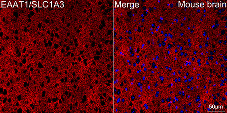
  • Confocal imaging of paraffin-embedded Mouse brain tissue using EAAT1/SLC1A3 Rabbit monoclonal antibody (STJ11102531, dilution 1:200) followed by a further incubation with Cy3 Goat Anti-Rabbit IgG (H+L) ( dilution 1:500) (Red). DAPI was used for nuclear staining (Blue). Objective: 40x. Perform microwave antigen retrieval with 0. 01 M citrate buffer (pH 6. 0) prior to IF staining.
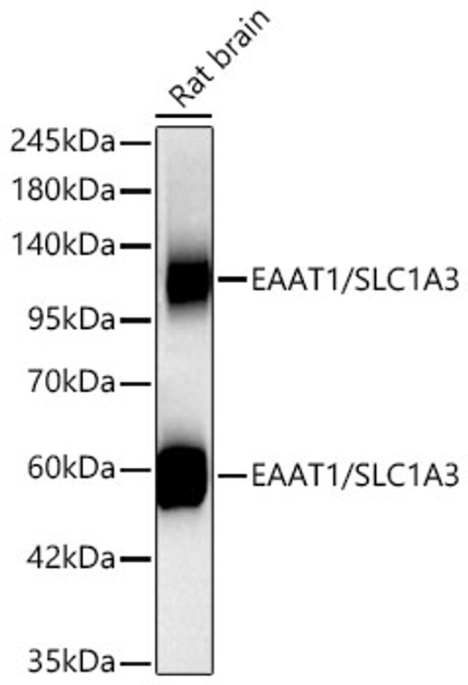
  • Western blot analysis of extracts of Rat brain, using EAAT1/SLC1A3 rabbit monoclonal antibody (STJ11102531) at 1:500 dilution. Secondary antibody: HRP Goat Anti-rabbit IgG (H+L) at 1:10000 dilution. Lysates/proteins: 25ug per lane. Blocking buffer: 3% non-fat dry milk in TBST. Detection: ECL Enhanced Kit. Exposure time: 3min.
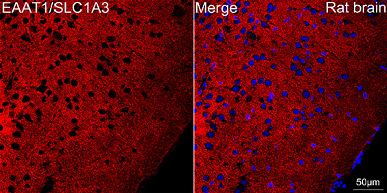
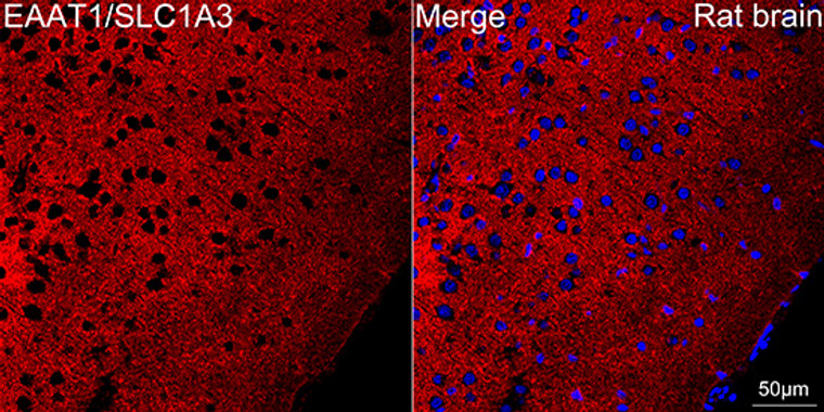
  • Confocal imaging of paraffin-embedded Rat brain tissue using EAAT1/SLC1A3 Rabbit monoclonal antibody (STJ11102531, dilution 1:200) followed by a further incubation with Cy3 Goat Anti-Rabbit IgG (H+L) ( dilution 1:500) (Red). DAPI was used for nuclear staining (Blue). Objective: 40x. Perform microwave antigen retrieval with 0. 01 M citrate buffer (pH 6. 0) prior to IF staining.
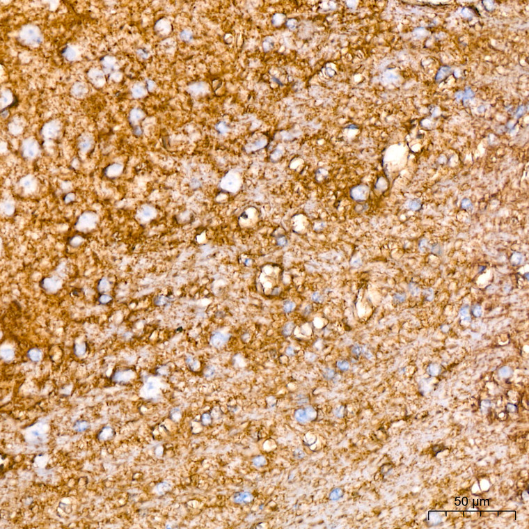
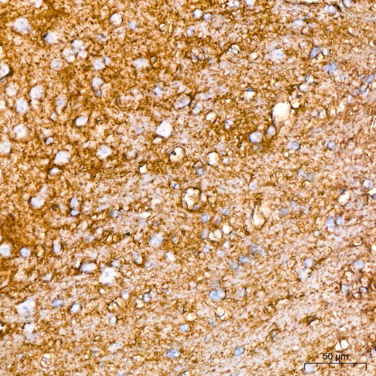
  • Immunohistochemistry of paraffin-embedded rat brain using EAAT1/SLC1A3 rabbit monoclonal antibody (STJ11102531) at dilution of 1:100 (40x lens).
  • Immunohistochemistry analysis of EAAT1/SLC1A3 in paraffin-embedded rat brain tissue using EAAT1/SLC1A3 Rabbit monoclonal antibody (STJ11102531) at a dilution of 1:200 (40x lens). High pressure antigen retrieval was performed with 0. 01 M citrate buffer (pH 6. 0) prior to immunohistochemistry staining.
  • Immunohistochemistry of paraffin-embedded human brain using EAAT1/SLC1A3 rabbit monoclonal antibody (STJ11102531) at dilution of 1:100 (40x lens).
  • Immunohistochemistry analysis of EAAT1/SLC1A3 in paraffin-embedded mouse brain tissue using EAAT1/SLC1A3 Rabbit monoclonal antibody (STJ11102531) at a dilution of 1:200 (40x lens). High pressure antigen retrieval was performed with 0. 01 M citrate buffer (pH 6. 0) prior to immunohistochemistry staining.
  • Immunohistochemistry of paraffin-embedded mouse spinal cord using EAAT1/SLC1A3 rabbit monoclonal antibody (STJ11102531) at dilution of 1:100 (40x lens).
  • Western blot analysis of lysates from Rat brain, using EAAT1/SLC1A3 Rabbit monoclonal antibody (STJ11102531) at 1:500 dilution. Secondary antibody: HRP Goat Anti-Rabbit IgG (H+L) (STJS000856) at 1:10000 dilution. Lysates/proteins: 25 Mu g per lane. Blocking buffer: 3% nonfat dry milk in TBST. Detection: ECL Enhanced Kit. Exposure time: 3min.
  • Western blot analysis of various lysates from Mouse brain using EAAT1/SLC1A3 Rabbit monoclonal antibody (STJ11102531) at 1:1000 dilution. Secondary antibody: HRP Goat Anti-Rabbit IgG (H+L) (STJS000856) at 1:10000 dilution. Lysates/proteins: 25 Mu g per lane. Blocking buffer: 3% nonfat dry milk in TBST. Detection: ECL Enhanced Kit. Exposure time: 3min.

Anti-SLC1A3 antibody (150-250) [S1MR] (STJ11102531)

SKU:
STJ11102531

Shipping:
Free Shipping
Current Stock:
Host: Rabbit
Applications: WB/IHC
Reactivity: Human/Mouse/Rat
Note: STRICTLY FOR FURTHER SCIENTIFIC RESEARCH USE ONLY (RUO). MUST NOT TO BE USED IN DIAGNOSTIC OR THERAPEUTIC APPLICATIONS.
Short Description: Rabbit monoclonal antibody anti-EAAT1 (150-250) is suitable for use in Western Blot and Immunohistochemistry research applications.
Clonality: Monoclonal
Clone ID: S1MR
Conjugation: Unconjugated
Isotype: IgG
Formulation: PBS with 0.02% Sodium Azide, 0.05% BSA, 50% Glycerol, pH7.3.
Purification: Affinity purification
Dilution Range: WB 1:500-1:1000
IHC-P 1:50-1:200
Storage Instruction: Store at-20°C for up to 1 year from the date of receipt, and avoid repeat freeze-thaw cycles.
Gene Symbol: SLC1A3
Gene ID: 6507
Uniprot ID: EAA1_HUMAN
Immunogen Region: 150-250
Immunogen: A synthetic peptide corresponding to a sequence within amino acids 150-250 of human EAAT1/SLC1A3 (P43003).
Immunogen Sequence: GTKENMHREGKIVRVTAADA FLDLIRNMFPPNLVEACFKQ FKTNYEKRSFKVPIQANETL VGAVINNVSEAMETLTRITE ELVPVPGSVNGVNALGLVVF S
Tissue Specificity Detected in brain. Detected at very much lower levels in heart, lung, placenta and skeletal muscle. Highly expressed in cerebellum, but also found in frontal cortex, hippocampus and basal ganglia.
Post Translational Modifications Glycosylated.
Function Sodium-dependent, high-affinity amino acid transporter that mediates the uptake of L-glutamate and also L-aspartate and D-aspartate. Functions as a symporter that transports one amino acid molecule together with two or three Na(+) ions and one proton, in parallel with the counter-transport of one K(+) ion. Mediates Cl(-) flux that is not coupled to amino acid transport.this avoids the accumulation of negative charges due to aspartate and Na(+) symport. Plays a redundant role in the rapid removal of released glutamate from the synaptic cleft, which is essential for terminating the postsynaptic action of glutamate.
Protein Name Excitatory Amino Acid Transporter 1
Sodium-Dependent Glutamate/Aspartate Transporter 1
Glast-1
Solute Carrier Family 1 Member 3
Database Links Reactome: R-HSA-210455
Reactome: R-HSA-210500
Reactome: R-HSA-425393
Reactome: R-HSA-5619062
Cellular Localisation Cell Membrane
Multi-Pass Membrane Protein
Alternative Antibody Names Anti-Excitatory Amino Acid Transporter 1 antibody
Anti-Sodium-Dependent Glutamate/Aspartate Transporter 1 antibody
Anti-Glast-1 antibody
Anti-Solute Carrier Family 1 Member 3 antibody
Anti-SLC1A3 antibody
Anti-EAAT1 antibody
Anti-GLAST antibody
Anti-GLAST1 antibody

Information sourced from Uniprot.org

12 months for antibodies. 6 months for ELISA Kits. Please see website T&Cs for further guidance