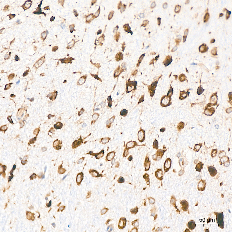
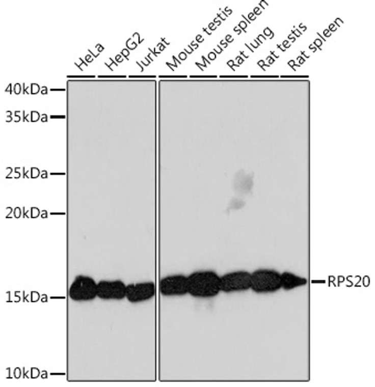
• Western blot analysis of extracts of various cell lines, using RPS20 rabbit monoclonal antibody (STJ11102489) at 1:1000 dilution. Secondary antibody: HRP Goat Anti-rabbit IgG (H+L) at 1:10000 dilution. Lysates/proteins: 25ug per lane. Blocking buffer: 3% non-fat dry milk in TBST. Detection: ECL Basic Kit. Exposure time: 3min.
  • Immunoprecipitation of RPS20 in 300 µg extracts from Hep G2 cells using 3 µg RPS20 Rabbit monoclonal antibody (STJ11102489). Western blot analysis was performed using RPS20 Rabbit monoclonal antibody (STJ11102489) at 1:1000 dilution.
  • Immunohistochemistry of paraffin-embedded rat brain using RPS20 rabbit monoclonal antibody (STJ11102489) at dilution of 1:100 (40x lens).
  • Immunofluorescence analysis of C6 cells using RPS20 Rabbit monoclonal antibody (STJ11102489) at dilution of 1:100 (40x lens). Secondary antibody: Cy3 Goat Anti-Rabbit IgG (H+L) at 1:500 dilution. Blue: DAPI for nuclear staining.
  • Immunohistochemistry of paraffin-embedded human colon using RPS20 rabbit monoclonal antibody (STJ11102489) at dilution of 1:100 (40x lens).
  • Immunohistochemistry analysis of RPS20 in paraffin-embedded rat colon tissue using RPS20 Rabbit monoclonal antibody (STJ11102489) at a dilution of 1:800 (40x lens). High pressure antigen retrieval was performed with 0. 01 M citrate buffer (pH 6. 0) prior to immunohistochemistry staining.
  • Immunohistochemistry of paraffin-embedded mouse brain using RPS20 rabbit monoclonal antibody (STJ11102489) at dilution of 1:100 (40x lens).
  • Immunohistochemistry analysis of RPS20 in paraffin-embedded rat brain tissue using RPS20 Rabbit monoclonal antibody (STJ11102489) at a dilution of 1:800 (40x lens). High pressure antigen retrieval was performed with 0. 01 M citrate buffer (pH 6. 0) prior to immunohistochemistry staining.
  • Immunofluorescence analysis of C6 cells using RPS20 rabbit monoclonal antibody (STJ11102489) at dilution of 1:100 (40x lens). Blue: DAPI for nuclear staining.
  • Immunohistochemistry analysis of RPS20 in paraffin-embedded mouse testis tissue using RPS20 Rabbit monoclonal antibody (STJ11102489) at a dilution of 1:800 (40x lens). High pressure antigen retrieval was performed with 0. 01 M citrate buffer (pH 6. 0) prior to immunohistochemistry staining.
  • Immunohistochemistry analysis of RPS20 in paraffin-embedded mouse brain tissue using RPS20 Rabbit monoclonal antibody (STJ11102489) at a dilution of 1:800 (40x lens). High pressure antigen retrieval was performed with 0. 01 M citrate buffer (pH 6. 0) prior to immunohistochemistry staining.
  • Immunohistochemistry analysis of RPS20 in paraffin-embedded human colon carcinoma tissue using RPS20 Rabbit monoclonal antibody (STJ11102489) at a dilution of 1:800 (40x lens). High pressure antigen retrieval was performed with 0. 01 M citrate buffer (pH 6. 0) prior to immunohistochemistry staining.
  • Western blot analysis of various lysates using RPS20 Rabbit monoclonal antibody (STJ11102489) at 1:1000 dilution. Secondary antibody: HRP Goat Anti-Rabbit IgG (H+L) (STJS000856) at 1:10000 dilution. Lysates/proteins: 25 Mu g per lane. Blocking buffer: 3% nonfat dry milk in TBST. Detection: ECL Basic Kit. Exposure time: 3min.

Anti-RPS20 antibody (1-100) [S9MR] (STJ11102489)

SKU:
STJ11102489

Shipping:
Free Shipping
Current Stock:
Host: Rabbit
Applications: WB/IHC/IF
Reactivity: Human/Mouse/Rat
Note: STRICTLY FOR FURTHER SCIENTIFIC RESEARCH USE ONLY (RUO). MUST NOT TO BE USED IN DIAGNOSTIC OR THERAPEUTIC APPLICATIONS.
Short Description: Rabbit monoclonal antibody anti-RPS20 (1-100) is suitable for use in Western Blot, Immunohistochemistry and Immunofluorescence research applications.
Clonality: Monoclonal
Clone ID: S9MR
Conjugation: Unconjugated
Isotype: IgG
Formulation: PBS with 0.02% Sodium Azide, 0.05% BSA, 50% Glycerol, pH7.3.
Purification: Affinity purification
Dilution Range: WB 1:500-1:1000
IHC-P 1:50-1:200
IF/ICC 1:50-1:200
Storage Instruction: Store at-20°C for up to 1 year from the date of receipt, and avoid repeat freeze-thaw cycles.
Gene Symbol: RPS20
Gene ID: 6224
Uniprot ID: RS20_HUMAN
Immunogen Region: 1-100
Immunogen: A synthetic peptide corresponding to a sequence within amino acids 1-100 of human RPS20 (P60866).
Immunogen Sequence: MAFKDTGKTPVEPEVAIHRI RITLTSRNVKSLEKVCADLI RGAKEKNLKVKGPVRMPTKT LRITTRKTPCGEGSKTWDRF QMRIHKRLIDLHSPSEIVKQ
Post Translational Modifications Polyubiquitinated by ZNF598 via 'Lys-63'-linked ubiquitin chains when a ribosome has stalled, initiating the ribosome quality control (RQC) pathway to degrade the potentially detrimental aberrant nascent polypeptide. Deubiquitinated by OTUD3 and USP21, antagonizing ZNF598 activity. Ufmylated by UFL1.
Function Component of the small ribosomal subunit. The ribosome is a large ribonucleoprotein complex responsible for the synthesis of proteins in the cell.
Protein Name Small Ribosomal Subunit Protein Us10
40s Ribosomal Protein S20
Database Links Reactome: R-HSA-156827
Reactome: R-HSA-156902
Reactome: R-HSA-1799339
Reactome: R-HSA-192823
Reactome: R-HSA-2408557
Reactome: R-HSA-6791226
Reactome: R-HSA-72649
Reactome: R-HSA-72689
Reactome: R-HSA-72695
Reactome: R-HSA-72702
Reactome: R-HSA-72706
Reactome: R-HSA-72764
Reactome: R-HSA-9010553
Reactome: R-HSA-9633012
Reactome: R-HSA-9735869
Reactome: R-HSA-9754678
Reactome: R-HSA-975956
Reactome: R-HSA-975957
Cellular Localisation Cytoplasm
Alternative Antibody Names Anti-Small Ribosomal Subunit Protein Us10 antibody
Anti-40s Ribosomal Protein S20 antibody
Anti-RPS20 antibody

Information sourced from Uniprot.org

12 months for antibodies. 6 months for ELISA Kits. Please see website T&Cs for further guidance