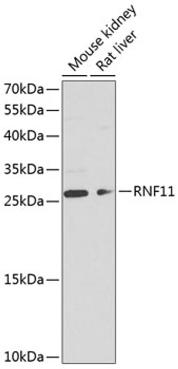
| Host: |
Rabbit |
| Applications: |
WB |
| Reactivity: |
Mouse/Rat |
| Note: |
STRICTLY FOR FURTHER SCIENTIFIC RESEARCH USE ONLY (RUO). MUST NOT TO BE USED IN DIAGNOSTIC OR THERAPEUTIC APPLICATIONS. |
| Short Description: |
Rabbit polyclonal antibody anti-RNF11 (1-154) is suitable for use in Western Blot research applications. |
| Clonality: |
Polyclonal |
| Conjugation: |
Unconjugated |
| Isotype: |
IgG |
| Formulation: |
PBS with 0.02% Sodium Azide, 50% Glycerol, pH7.3. |
| Purification: |
Affinity purification |
| Dilution Range: |
WB 1:500-1:2000 |
| Storage Instruction: |
Store at-20°C for up to 1 year from the date of receipt, and avoid repeat freeze-thaw cycles. |
| Gene Symbol: |
RNF11 |
| Gene ID: |
26994 |
| Uniprot ID: |
RNF11_HUMAN |
| Immunogen Region: |
1-154 |
| Immunogen: |
Recombinant fusion protein containing a sequence corresponding to amino acids 1-154 of human RNF11 (NP_055187.1). |
| Immunogen Sequence: |
MGNCLKSPTSDDISLLHESQ SDRASFGEGTEPDQEPPPPY QEQVPVPVYHPTPSQTRLAT QLTEEEQIRIAQRIGLIQHL PKGVYDPGRDGSEKKIRECV ICMMDFVYGDPIRFLPCMHI YHLDCIDDWLMRSFTCPSCM EPVDAALLSSYETN |
| Tissue Specificity | Expressed at low levels in the lung, liver, kidney, pancreas, spleen, prostate, thymus, ovary, small intestine, colon, and peripheral blood lymphocytes, and, at intermediate levels, in the testis, heart, brain and placenta. Highest expression in the skeletal muscle. In the brain, expressed at different levels in several regions: high levels in the amygdala, moderate in the hippocampus and thalamus, low in the caudate and extremely low levels in the corpus callosum (at protein level). Restricted to neurons, enriched in somatodendritic compartments and excluded from white matter (at protein level). In substantia nigra, present in cell bodies and processes of dopaminergic and nondopaminergic cells (at protein level). In Parkinson disease, sequestered in Lewy bodies and neurites. Overexpressed in breast cancer cells, but not detected in the surrounding stroma and weakly, if at all, in normal breast epithelial cells (at protein level). Also expressed in several tumor cell lines. |
| Post Translational Modifications | Ubiquitinated in the presence of ITCH, or SMURF2, and UBE2D1, as well as WWP1. Phosphorylation by PKB/AKT1 may accelerate degradation by the proteasome. Acylation at both Gly-2 and Cys-4 is required for proper localization to the endosomes. |
| Function | Essential component of a ubiquitin-editing protein complex, comprising also TNFAIP3, ITCH and TAX1BP1, that ensures the transient nature of inflammatory signaling pathways. Promotes the association of TNFAIP3 to RIPK1 after TNF stimulation. TNFAIP3 deubiquitinates 'Lys-63' polyubiquitin chains on RIPK1 and catalyzes the formation of 'Lys-48'-polyubiquitin chains. This leads to RIPK1 proteasomal degradation and consequently termination of the TNF- or LPS-mediated activation of NF-kappa-B. Recruits STAMBP to the E3 ubiquitin-ligase SMURF2 for ubiquitination, leading to its degradation by the 26S proteasome. |
| Protein Name | Ring Finger Protein 11 |
| Cellular Localisation | Early EndosomeRecycling EndosomeCytoplasmNucleusPredominantly CytoplasmicWhen UnphosphorylatedAnd NuclearWhen Phosphorylated By Pkb/Akt1 |
| Alternative Antibody Names | Anti-Ring Finger Protein 11 antibodyAnti-RNF11 antibodyAnti-CGI-123 antibody |
Information sourced from Uniprot.org
12 months for antibodies. 6 months for ELISA Kits. Please see website T&Cs for further guidance


