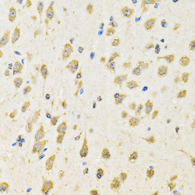
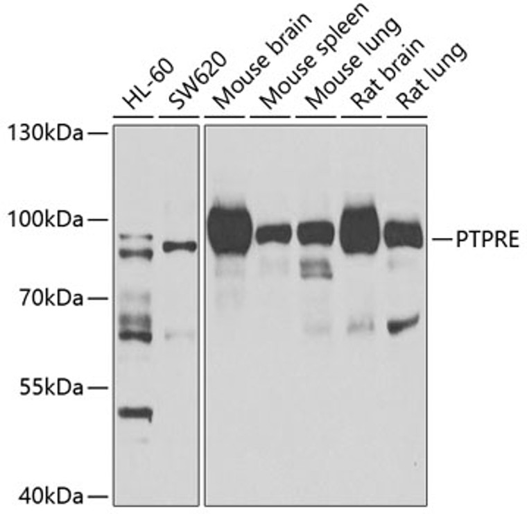
| Host: |
Rabbit |
| Applications: |
WB/IHC |
| Reactivity: |
Human/Mouse/Rat |
| Note: |
STRICTLY FOR FURTHER SCIENTIFIC RESEARCH USE ONLY (RUO). MUST NOT TO BE USED IN DIAGNOSTIC OR THERAPEUTIC APPLICATIONS. |
| Short Description: |
Rabbit polyclonal antibody anti-PTPRE (20-200) is suitable for use in Western Blot and Immunohistochemistry research applications. |
| Clonality: |
Polyclonal |
| Conjugation: |
Unconjugated |
| Isotype: |
IgG |
| Formulation: |
PBS with 0.02% Sodium Azide, 50% Glycerol, pH7.3. |
| Purification: |
Affinity purification |
| Dilution Range: |
WB 1:500-1:2000IHC-P 1:50-1:200 |
| Storage Instruction: |
Store at-20°C for up to 1 year from the date of receipt, and avoid repeat freeze-thaw cycles. |
| Gene Symbol: |
PTPRE |
| Gene ID: |
5791 |
| Uniprot ID: |
PTPRE_HUMAN |
| Immunogen Region: |
20-200 |
| Immunogen: |
Recombinant fusion protein containing a sequence corresponding to amino acids 20-200 of human PTPRE (NP_006495.1). |
| Immunogen Sequence: |
LRGNETTADSNETTTTSGPP DPGASQPLLAWLLLPLLLLL LVLLLAAYFFRFRKQRKAVV STSDKKMPNGILEEQEQQRV MLLSRSPSGPKKYFPIPVEH LEEEIRIRSADDCKQFREEF NSLPSGHIQGTFELANKEEN REKNRYPNILPNDHSRVILS QLDGIPCSDYINASYIDGYK E |
| Tissue Specificity | Expressed in giant cell tumor (osteoclastoma rich in multinucleated osteoclastic cells). |
| Post Translational Modifications | A catalytically active cytoplasmic form (p65) is produced by proteolytic cleavage of either isoform 1, isoform 2 or isoform 3. Isoform 1 and isoform 2 are phosphorylated on tyrosine residues by tyrosine kinase Neu. Isoform 1 is glycosylated. |
| Function | Isoform 1 plays a critical role in signaling transduction pathways and phosphoprotein network topology in red blood cells. May play a role in osteoclast formation and function. Isoform 2 acts as a negative regulator of insulin receptor (IR) signaling in skeletal muscle. Regulates insulin-induced tyrosine phosphorylation of insulin receptor (IR) and insulin receptor substrate 1 (IRS-1), phosphorylation of protein kinase B and glycogen synthase kinase-3 and insulin induced stimulation of glucose uptake. Isoform 1 and isoform 2 act as a negative regulator of FceRI-mediated signal transduction leading to cytokine production and degranulation, most likely by acting at the level of SYK to affect downstream events such as phosphorylation of SLP76 and LAT and mobilization of Ca(2+). |
| Protein Name | Receptor-Type Tyrosine-Protein Phosphatase EpsilonProtein-Tyrosine Phosphatase EpsilonR-Ptp-Epsilon |
| Cellular Localisation | Isoform 1: Cell MembraneSingle-Pass Type I Membrane ProteinIsoform 2: CytoplasmPredominantly CytoplasmicA Small Fraction Is Also Associated With Nucleus And MembraneInsulin Induces Translocation To The MembraneIsoform 3: Cytoplasm |
| Alternative Antibody Names | Anti-Receptor-Type Tyrosine-Protein Phosphatase Epsilon antibodyAnti-Protein-Tyrosine Phosphatase Epsilon antibodyAnti-R-Ptp-Epsilon antibodyAnti-PTPRE antibody |
Information sourced from Uniprot.org
12 months for antibodies. 6 months for ELISA Kits. Please see website T&Cs for further guidance


