| Host: |
Rabbit |
| Applications: |
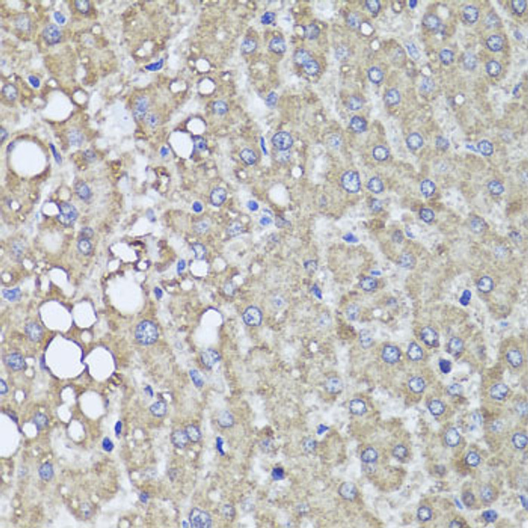
WB/IHC-P/IF/ICC/IP/ELISA |
| Reactivity: |
Human/Mouse/Rat |
| Note: |
STRICTLY FOR FURTHER SCIENTIFIC RESEARCH USE ONLY (RUO). MUST NOT TO BE USED IN DIAGNOSTIC OR THERAPEUTIC APPLICATIONS. |
| Clonality: |
Polyclonal |
| Conjugation: |
Unconjugated |
| Isotype: |
IgG |
| Formulation: |
PBS with 0.02% Sodium Azide, 50% Glycerol, pH 7.3. |
| Purification: |
Affinity purification |
| Concentration: |
Lot specific |
| Dilution Range: |
WB:1:500-1:2000IHC-P:1:50-1:100IF/ICC:1:50-1:100IP:1:50-1:100ELISA:Recommended starting concentration is 1 Mu g/mL. Please optimize the concentration based on your specific assay requirements. |
| Storage Instruction: |
Store at-20°C for up to 1 year from the date of receipt, and avoid repeat freeze-thaw cycles. |
| Gene Symbol: |
PIK3C2A |
| Gene ID: |
5286 |
| Uniprot ID: |
P3C2A_HUMAN |
| Immunogen Region: |
1-200 |
| Specificity: |
Recombinant fusion protein containing a sequence corresponding to amino acids 1-200 of human PIK3C2A (NP_002636.2). |
| Immunogen Sequence: |
MAQISSNSGFKECPSSHPEP TRAKDVDKEEALQMEAEALA KLQKDRQVTDNQRGFELSSS TRKKAQVYNKQDYDLMVFPE SDSQKRALDIDVEKLTQAEL EKLLLDDSFETKKTPVLPVT PILSPSFSAQLYFRPTIQRG QWPPGLPGPSTYALPSIYPS TYSKQAAFQNGFNPRMPTFP STEPIYLSLPGQSPYFSYPL |
| Tissue Specificity | Expressed in columnar and transitional epithelia, mononuclear cells, smooth muscle cells, and endothelial cells lining capillaries and small venules (at protein level). Ubiquitously expressed, with highest levels in heart, placenta and ovary, and lowest levels in the kidney. Detected at low levels in islets of Langerhans from type 2 diabetes mellitus individuals. |
| Post Translational Modifications | Phosphorylated upon insulin stimulation.which may lead to enzyme activation. Phosphorylated on Ser-259 during mitosis and upon UV irradiation.which does not change enzymatic activity but leads to proteasomal degradation. Ser-259 phosphorylation may be mediated by CDK1 or JNK, depending on the physiological state of the cell. |
| Function | Generates phosphatidylinositol 3-phosphate (PtdIns3P) and phosphatidylinositol 3,4-bisphosphate (PtdIns(3,4)P2) that act as second messengers. Has a role in several intracellular trafficking events. Functions in insulin signaling and secretion. Required for translocation of the glucose transporter SLC2A4/GLUT4 to the plasma membrane and glucose uptake in response to insulin-mediated RHOQ activation. Regulates insulin secretion through two different mechanisms: involved in glucose-induced insulin secretion downstream of insulin receptor in a pathway that involves AKT1 activation and TBC1D4/AS160 phosphorylation, and participates in the late step of insulin granule exocytosis probably in insulin granule fusion. Synthesizes PtdIns3P in response to insulin signaling. Functions in clathrin-coated endocytic vesicle formation and distribution. Regulates dynamin-independent endocytosis, probably by recruiting EEA1 to internalizing vesicles. In neurosecretory cells synthesizes PtdIns3P on large dense core vesicles. Participates in calcium induced contraction of vascular smooth muscle by regulating myosin light chain (MLC) phosphorylation through a mechanism involving Rho kinase-dependent phosphorylation of the MLCP-regulatory subunit MYPT1. May play a role in the EGF signaling cascade. May be involved in mitosis and UV-induced damage response. Required for maintenance of normal renal structure and function by supporting normal podocyte function. Involved in the regulation of ciliogenesis and trafficking of ciliary components. |
| Protein Name | Phosphatidylinositol 4-Phosphate 3-Kinase C2 Domain-Containing Subunit AlphaPi3k-C2-AlphaPtdins-3-Kinase C2 Subunit Alpha Phosphoinositide 3-Kinase-C2-Alpha |
| Database Links | Reactome: R-HSA-1660499Reactome: R-HSA-1660514Reactome: R-HSA-1660516Reactome: R-HSA-1660517Reactome: R-HSA-432722Reactome: R-HSA-8856828 |
| Cellular Localisation | Cell MembraneCytoplasmic VesicleClathrin-Coated VesicleNucleusCytoplasmGolgi ApparatusTrans-Golgi NetworkInserts Preferentially Into Membranes Containing Ptdins(45)P2Associated With Rna-Containing Structures |
| Alternative Antibody Names | Anti-Phosphatidylinositol 4-Phosphate 3-Kinase C2 Domain-Containing Subunit Alpha antibodyAnti-Pi3k-C2-Alpha antibodyAnti-Ptdins-3-Kinase C2 Subunit Alpha antibodyAnti-Phosphoinositide 3-Kinase-C2-Alpha antibodyAnti-PIK3C2A antibody |
Information sourced from Uniprot.org
12 months for antibodies. 6 months for ELISA Kits. Please see website T&Cs for further guidance


