| Tissue Specificity | Expressed in pyramidal neurons in all cortical laminae of the visual cortex, in neurons of the substantia nigra pars compacta and caudate putamen (at protein level). Expressed in neutrophils (at protein level). Expressed in the brain. Expressed throughout the adult brain, but at a lower level than in heart and liver. Also expressed in placenta, lung, skeletal muscle, kidney and pancreas. In the brain, expressed in the cerebellum, cerebral cortex, medulla, spinal cord occipital pole, frontal lobe, temporal lobe and putamen. Expression is particularly high in brain dopaminoceptive areas. |
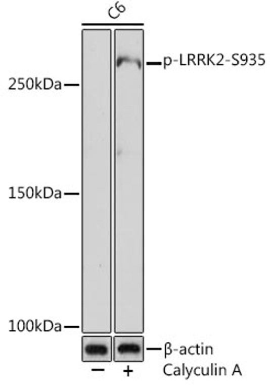
| Post Translational Modifications | Autophosphorylated. Phosphorylation of Ser-910 and either Ser-935 or Ser-1444 facilitates interaction with YWHAG. Phosphorylation of Ser-910 and/or Ser-935 facilitates interaction with SFN. Ubiquitinated by TRIM1.undergoes 'Lys-48'-linked polyubiquitination leading to proteasomal degradation. |
| Function | Serine/threonine-protein kinase which phosphorylates a broad range of proteins involved in multiple processes such as neuronal plasticity, innate immunity, autophagy, and vesicle trafficking. Is a key regulator of RAB GTPases by regulating the GTP/GDP exchange and interaction partners of RABs through phosphorylation. Phosphorylates RAB3A, RAB3B, RAB3C, RAB3D, RAB5A, RAB5B, RAB5C, RAB8A, RAB8B, RAB10, RAB12, RAB35, and RAB43. Regulates the RAB3IP-catalyzed GDP/GTP exchange for RAB8A through the phosphorylation of 'Thr-72' on RAB8A. Inhibits the interaction between RAB8A and GDI1 and/or GDI2 by phosphorylating 'Thr-72' on RAB8A. Regulates primary ciliogenesis through phosphorylation of RAB8A and RAB10, which promotes SHH signaling in the brain. Together with RAB29, plays a role in the retrograde trafficking pathway for recycling proteins, such as mannose-6-phosphate receptor (M6PR), between lysosomes and the Golgi apparatus in a retromer-dependent manner. Regulates neuronal process morphology in the intact central nervous system (CNS). Plays a role in synaptic vesicle trafficking. Plays an important role in recruiting SEC16A to endoplasmic reticulum exit sites (ERES) and in regulating ER to Golgi vesicle-mediated transport and ERES organization. Positively regulates autophagy through a calcium-dependent activation of the CaMKK/AMPK signaling pathway. The process involves activation of nicotinic acid adenine dinucleotide phosphate (NAADP) receptors, increase in lysosomal pH, and calcium release from lysosomes. Phosphorylates PRDX3. By phosphorylating APP on 'Thr-743', which promotes the production and the nuclear translocation of the APP intracellular domain (AICD), regulates dopaminergic neuron apoptosis. Acts as a positive regulator of innate immunity by mediating phosphorylation of RIPK2 downstream of NOD1 and NOD2, thereby enhancing RIPK2 activation. Independent of its kinase activity, inhibits the proteasomal degradation of MAPT, thus promoting MAPT oligomerization and secretion. In addition, has GTPase activity via its Roc domain which regulates LRRK2 kinase activity. |
| Protein Name | Leucine-Rich Repeat Serine/Threonine-Protein Kinase 2Dardarin |
| Database Links | Reactome: R-HSA-8857538 |
| Cellular Localisation | Cytoplasmic VesiclePerikaryonGolgi Apparatus MembranePeripheral Membrane ProteinCell ProjectionAxonDendriteEndoplasmic Reticulum MembraneSecretory VesicleSynaptic Vesicle MembraneEndosomeLysosomeMitochondrion Outer MembraneCytoplasmCytoskeletonColocalized With Rab29 Along Tubular Structures Emerging From Golgi ApparatusLocalizes To Endoplasmic Reticulum Exit Sites (Eres)Also Known As Transitional Endoplasmic Reticulum (Ter) |
| Alternative Antibody Names | Anti-Leucine-Rich Repeat Serine/Threonine-Protein Kinase 2 antibodyAnti-Dardarin antibodyAnti-LRRK2 antibodyAnti-PARK8 antibody |


