| Host: |
Rabbit |
| Applications: |
WB |
| Reactivity: |
Human |
| Note: |
STRICTLY FOR FURTHER SCIENTIFIC RESEARCH USE ONLY (RUO). MUST NOT TO BE USED IN DIAGNOSTIC OR THERAPEUTIC APPLICATIONS. |
| Short Description: |
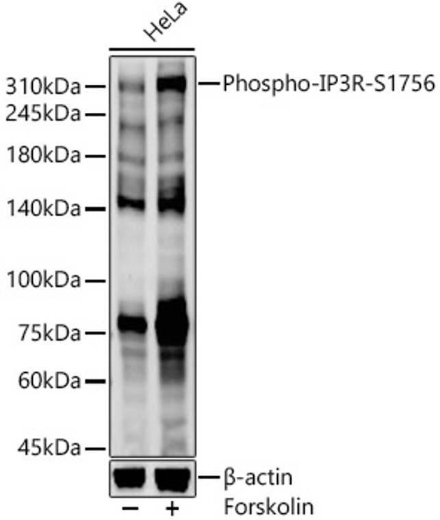
Rabbit polyclonal antibody anti-Phospho-Inositol 1-4-5-Trisphosphate Receptor Type 1-S1756 is suitable for use in Western Blot research applications. |
| Clonality: |
Polyclonal |
| Conjugation: |
Unconjugated |
| Isotype: |
IgG |
| Formulation: |
PBS with 0.01% thimerosal, 50% Glycerol, pH7.3. |
| Purification: |
Affinity purification |
| Dilution Range: |
WB 1:100-1:500 |
| Storage Instruction: |
Store at-20°C for up to 1 year from the date of receipt, and avoid repeat freeze-thaw cycles. |
| Gene Symbol: |
ITPR1 |
| Gene ID: |
3708 |
| Uniprot ID: |
ITPR1_HUMAN |
| Immunogen: |
A synthetic phosphorylated peptide around S1756 of human ITPR1 (XP_011531986.1). |
| Immunogen Sequence: |
RESLT |
| Tissue Specificity | Widely expressed. |
| Post Translational Modifications | Phosphorylated on tyrosine residues. Ubiquitination at multiple lysines targets ITPR1 for proteasomal degradation. Approximately 40% of the ITPR1-associated ubiquitin is monoubiquitin, and polyubiquitins are both 'Lys-48'- and 'Lys-63'-linked. Phosphorylated by cAMP kinase (PKA). Phosphorylation prevents the ligand-induced opening of the calcium channels. Phosphorylation by PKA increases the interaction with inositol 1,4,5-trisphosphate and decreases the interaction with AHCYL1. Palmitoylated by ZDHHC6 in immune cells, leading to regulation of ITPR1 stability and function. |
| Function | Intracellular channel that mediates calcium release from the endoplasmic reticulum following stimulation by inositol 1,4,5-trisphosphate. Involved in the regulation of epithelial secretion of electrolytes and fluid through the interaction with AHCYL1. Plays a role in ER stress-induced apoptosis. Cytoplasmic calcium released from the ER triggers apoptosis by the activation of CaM kinase II, eventually leading to the activation of downstream apoptosis pathways. |
| Protein Name | Inositol 1 -4 -5-Trisphosphate Receptor Type 1Ip3 Receptor Isoform 1Ip3r 1Insp3r1Type 1 Inositol 1 -4 -5-Trisphosphate ReceptorType 1 Insp3 Receptor |
| Database Links | Reactome: R-HSA-112043Reactome: R-HSA-114508Reactome: R-HSA-139853Reactome: R-HSA-1489509Reactome: R-HSA-2029485Reactome: R-HSA-2871809Reactome: R-HSA-381676Reactome: R-HSA-4086398Reactome: R-HSA-418457Reactome: R-HSA-422356Reactome: R-HSA-5218921Reactome: R-HSA-5578775Reactome: R-HSA-5607763Reactome: R-HSA-9664323Reactome: R-HSA-983695 |
| Cellular Localisation | Endoplasmic Reticulum MembraneMulti-Pass Membrane ProteinCytoplasmic VesicleSecretory Vesicle MembraneCytoplasmPerinuclear RegionEndoplasmic Reticulum And Secretory Granules |
| Alternative Antibody Names | Anti-Inositol 1 -4 -5-Trisphosphate Receptor Type 1 antibodyAnti-Ip3 Receptor Isoform 1 antibodyAnti-Ip3r 1 antibodyAnti-Insp3r1 antibodyAnti-Type 1 Inositol 1 -4 -5-Trisphosphate Receptor antibodyAnti-Type 1 Insp3 Receptor antibodyAnti-ITPR1 antibodyAnti-INSP3R1 antibody |
Information sourced from Uniprot.org
12 months for antibodies. 6 months for ELISA Kits. Please see website T&Cs for further guidance


