• Western blot analysis of extracts of various cell lines, using Phospho-Histone H3-T11 antibody (STJ22169) at 1:1000 dilution. HeLa cells, NIH/3T3 cells and C6 cells were treated by Nocodazole (50 ng/mL) at 37 °C for 20 hours. Secondary antibody: HRP Goat Anti-rabbit IgG (H+L) at 1:10000 dilution. Lysates/proteins: 25ug per lane. Blocking buffer: 3% BSA. Detection: ECL Basic Kit. Exposure time: 1s.
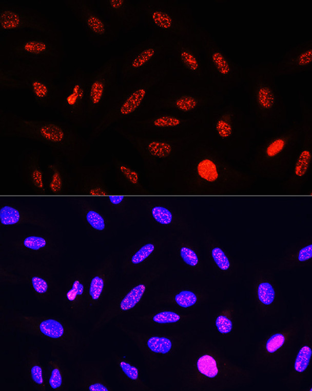
  • Immunofluorescence analysis of U-2 OS cells using Phospho-Histone H3-T11 Rabbit polyclonal antibody (STJ22169) at dilution of 100 (40x lens). Secondary antibody: Cy3 Goat Anti-Rabbit IgG (H+L) at 1:500 dilution. Blue: DAPI for nuclear staining.
  • Western blot analysis of extracts of NIH/3T3 cells, using phospho-STK4-T387 polyclonal antibody (STJ22169) at 1:1000 dilution or Histone H3 antibody (A15741). HeLa, NIH/3T3 and C6 cellls were treated by nocodazole (50 ng/mL) at 37 °C for 20 hours. Secondary antibody: HRP Goat Anti-rabbit IgG (H+L) at 1:10000 dilution. Lysates/proteins: 25ug per lane. Blocking buffer: 3% BSA. Detection: ECL Basic Kit. Exposure time: 1s.
  • Immunofluorescence analysis of NIH-3T3 cells using Phospho-Histone H3-T11 Rabbit polyclonal antibody (STJ22169) at dilution of 100 (40x lens). Secondary antibody: Cy3 Goat Anti-Rabbit IgG (H+L) at 1:500 dilution. Blue: DAPI for nuclear staining.
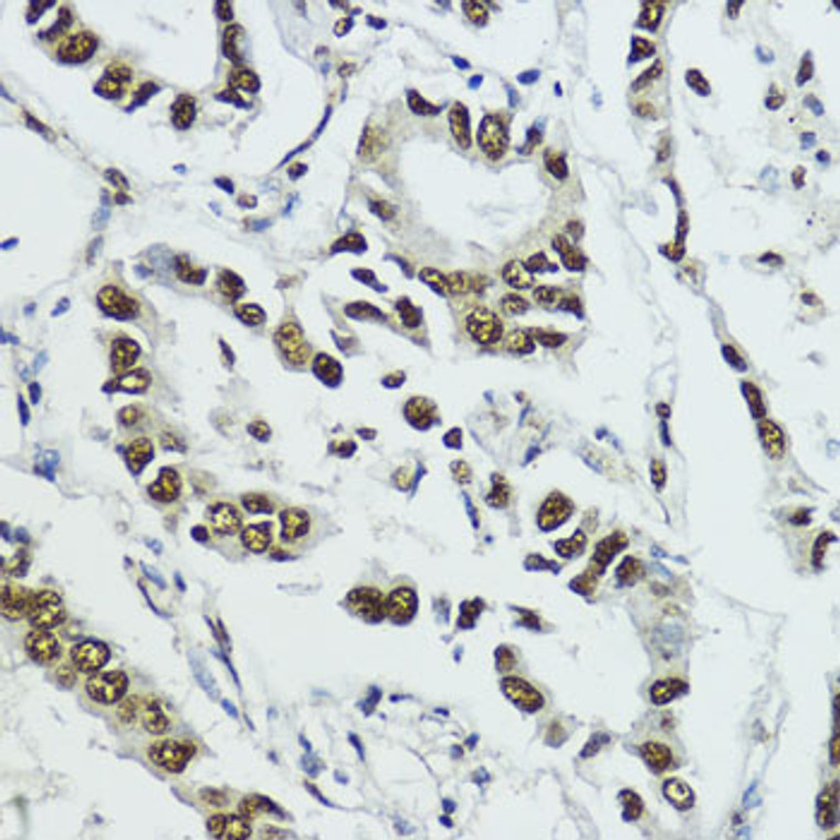
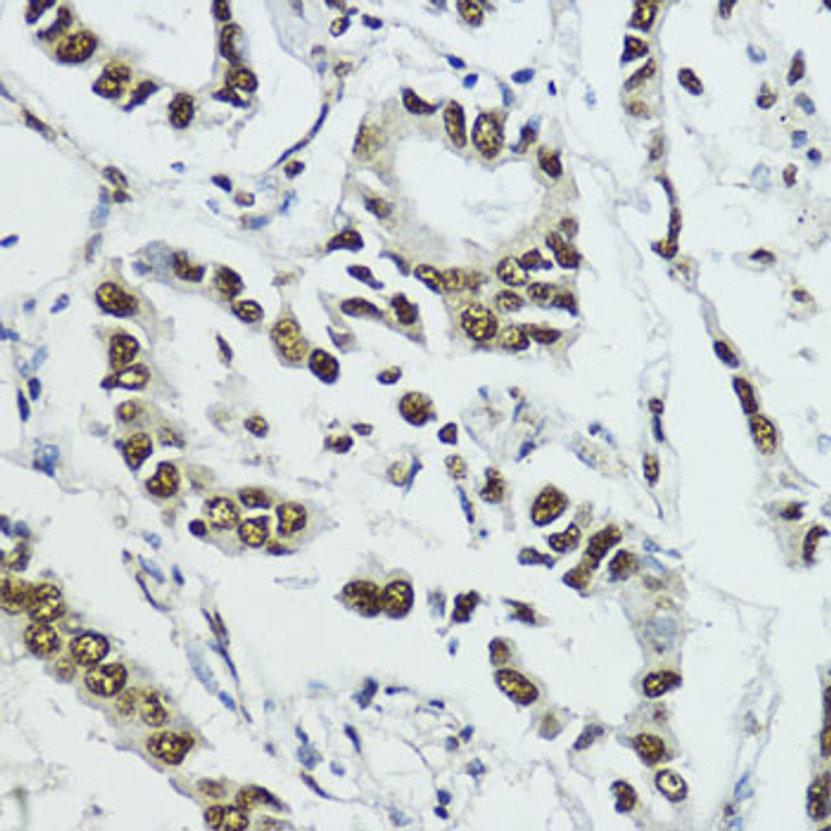
  • Immunohistochemistry of paraffin-embedded human lung cancer using Phospho-Histone H3-T11 antibody (STJ22169) at dilution of 1:100 (40x lens).
  • Immunofluorescence analysis of C6 cells using Phospho-Histone H3-T11 Rabbit polyclonal antibody (STJ22169) at dilution of 100 (40x lens). Secondary antibody: Cy3 Goat Anti-Rabbit IgG (H+L) at 1:500 dilution. Blue: DAPI for nuclear staining.
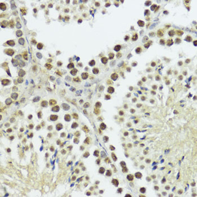
  • Immunohistochemistry of paraffin-embedded rat testis using Phospho-Histone H3-T11 antibody (STJ22169) at dilution of 1:100 (40x lens).
  • Immunohistochemistry analysis of Phospho-Histone H3-T11 in paraffin-embedded mouse testis using Phospho-Histone H3-T11 Rabbit polyclonal antibody (STJ22169) at dilution of 1:100 (40x lens). Perform microwave antigen retrieval with 10 mM Tris/EDTA buffer pH 9. 0 before commencing with immunohistochemistry staining protocol.
  • Immunohistochemistry of paraffin-embedded mouse testis using Phospho-Histone H3-T11 antibody (STJ22169) at dilution of 1:100 (40x lens).
  • Immunohistochemistry analysis of Phospho-Histone H3-T11 in paraffin-embedded rat testis using Phospho-Histone H3-T11 Rabbit polyclonal antibody (STJ22169) at dilution of 1:100 (40x lens). Perform microwave antigen retrieval with 10 mM Tris/EDTA buffer pH 9. 0 before commencing with immunohistochemistry staining protocol.
  • Immunofluorescence analysis of C6 cells using Phospho-Histone H3-T11 rabbit polyclonal antibody (STJ22169) at dilution of 100 (40x lens). Blue: DAPI for nuclear staining.
  • Immunohistochemistry analysis of Phospho-Histone H3-T11 in paraffin-embedded human lung cancer using Phospho-Histone H3-T11 Rabbit polyclonal antibody (STJ22169) at dilution of 1:100 (40x lens). Perform microwave antigen retrieval with 10 mM Tris/EDTA buffer pH 9. 0 before commencing with immunohistochemistry staining protocol.
  • Immunofluorescence analysis of NIH-3T3 cells using Phospho-Histone H3-T11 rabbit polyclonal antibody (STJ22169) at dilution of 100 (40x lens). Blue: DAPI for nuclear staining.
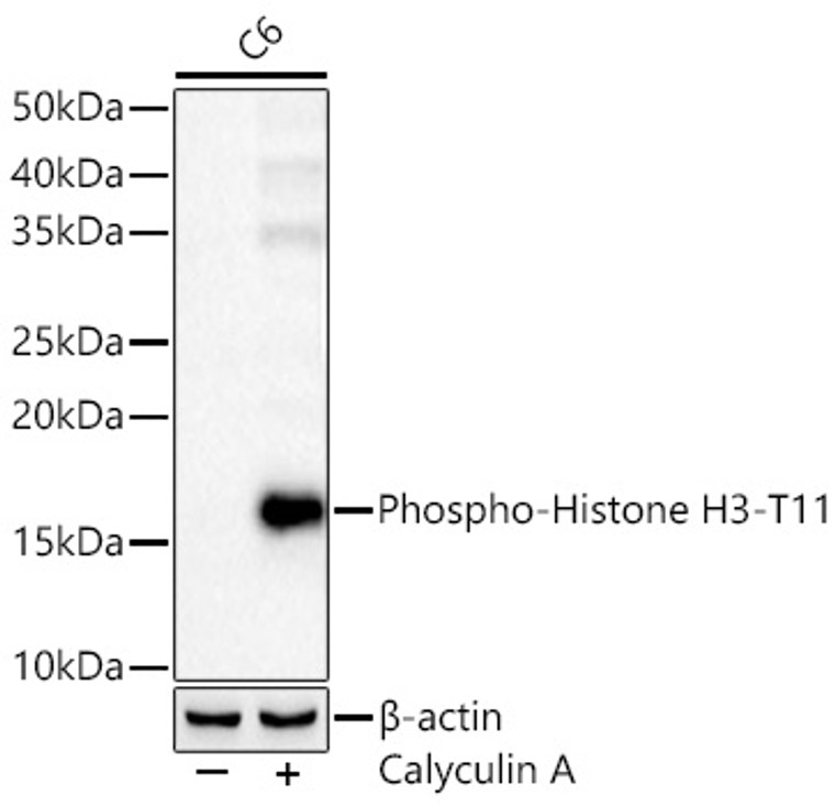
  • Western blot analysis of lysates from C6 cells using Phospho-Histone H3-T11 Rabbit polyclonal antibody (STJ22169) at 1:2000 dilution. C6 cells were treated by Calyculin A (100 nM) at 37 °C for 30 minutes after serum-starvation overnight. Secondary antibody:HRP Goat Anti-Rabbit IgG (H+L) (STJS000856) at 1:10000 dilution. Lysates/proteins: 25  Mu g per lane. Blocking buffer: 3% nonfat dry milk in TBST. Detection:ECL Basic Kit. Exposuretime: 60s.
  • Immunofluorescence analysis of U-2 OS cells using Phospho-Histone H3-T11 rabbit polyclonal antibody (STJ22169) at dilution of 100 (40x lens). Blue: DAPI for nuclear staining.

Anti-Phospho-Histone H3-T11 antibody (STJ22169)

SKU:
STJ22169

Shipping:
Free Shipping
Current Stock:
Host: Rabbit
Applications: WB/IHC/IF
Reactivity: Human/Mouse/Rat
Note: STRICTLY FOR FURTHER SCIENTIFIC RESEARCH USE ONLY (RUO). MUST NOT TO BE USED IN DIAGNOSTIC OR THERAPEUTIC APPLICATIONS.
Short Description: Rabbit polyclonal antibody anti-Phospho-Histone H3-T11 is suitable for use in Western Blot, Immunohistochemistry and Immunofluorescence research applications.
Clonality: Polyclonal
Conjugation: Unconjugated
Isotype: IgG
Formulation: PBS with 0.01% Thimerosal, 50% Glycerol, pH7.3.
Purification: Affinity purification
Dilution Range: WB 1:100-1:500
IHC-P 1:50-1:200
IF/ICC 1:50-1:200
Storage Instruction: Store at-20°C for up to 1 year from the date of receipt, and avoid repeat freeze-thaw cycles.
Immunogen: A synthetic phosphorylated peptide around T11 of human HIST1H3B (NP_003520.1).
Immunogen Sequence: KSTGG
Background The protein encoded by this gene belongs to the SMAD, a family of proteins similar to the gene products of the Drosophila gene mothers against decapentaplegic (Mad) and the C. elegans gene Sma. SMAD proteins are signal transducers and transcriptional modulators that mediate multiple signaling pathways. This protein mediates the signal of the transforming growth factor (TGF)-beta, and thus regulates multiple cellular processes, such as cell proliferation, apoptosis, and differentiation. This protein is recruited to the TGF-beta receptors through its interaction with the SMAD anchor for receptor activation (SARA) protein. In response to TGF-beta signal, this protein is phosphorylated by the TGF-beta receptors. The phosphorylation induces the dissociation of this protein with SARA and the association with the family member SMAD4. The association with SMAD4 is important for the translocation of this protein into the nucleus, where it binds to target promoters and forms a transcription repressor complex with other cofactors. This protein can also be phosphorylated by activin type 1 receptor kinase, and mediates the signal from the activin. Alternatively spliced transcript variants have been observed for this gene.

Information sourced from Uniprot.org

12 months for antibodies. 6 months for ELISA Kits. Please see website T&Cs for further guidance