• Immunohistochemical analysis of paraffin-embedded Mouse-lung tissue. 1, Akt (phospho Thr308) Polyclonal Antibody was diluted at 1:200 (4°C, overnight). 2, Sodium citrate pH 6.0 was used for antibody retrieval (>98°C, 20min). 3, Secondary antibody was diluted at 1:200 (room tempeRature, 30min). Negative control was used by secondary antibody only.
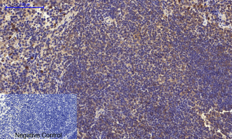
  • Immunohistochemical analysis of paraffin-embedded Mouse-spleen tissue. 1, Akt (phospho Thr308) Polyclonal Antibody was diluted at 1:200 (4°C, overnight). 2, Sodium citrate pH 6.0 was used for antibody retrieval (>98°C, 20min). 3, Secondary antibody was diluted at 1:200 (room tempeRature, 30min). Negative control was used by secondary antibody only.
  • Immunohistochemical analysis of paraffin-embedded Rat-spinal-cord tissue. 1, Akt (phospho Thr308) Polyclonal Antibody was diluted at 1:200 (4°C, overnight). 2, Sodium citrate pH 6.0 was used for antibody retrieval (>98°C, 20min). 3, Secondary antibody was diluted at 1:200 (room tempeRature, 30min). Negative control was used by secondary antibody only.
  • Immunohistochemical analysis of paraffin-embedded Rat-spleen tissue. 1, Akt (phospho Thr308) Polyclonal Antibody was diluted at 1:200 (4°C, overnight). 2, Sodium citrate pH 6.0 was used for antibody retrieval (>98°C, 20min). 3, Secondary antibody was diluted at 1:200 (room tempeRature, 30min). Negative control was used by secondary antibody only.
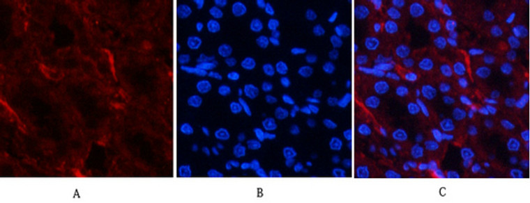
  • Immunofluorescence analysis of human-lung tissue. 1, Akt (phospho Thr308) Polyclonal Antibody (red) was diluted at 1:200 (4°C, overnight). 2, Cy3 labled Secondary antibody was diluted at 1:300 (room temperature, 50min).3, Picture B: DAPI (blue) 10min. Picture A:Target. Picture B: DAPI. Picture C: merge of A+B
  • Immunohistochemical analysis of paraffin-embedded Rat-lung tissue. 1, Akt (phospho Thr308) Polyclonal Antibody was diluted at 1:200 (4°C, overnight). 2, Sodium citrate pH 6.0 was used for antibody retrieval (>98°C, 20min). 3, Secondary antibody was diluted at 1:200 (room tempeRature, 30min). Negative control was used by secondary antibody only.
  • Immunohistochemical analysis of paraffin-embedded Human-colon tissue. 1, Akt (phospho Thr308) Polyclonal Antibody was diluted at 1:200 (4°C, overnight). 2, Sodium citrate pH 6.0 was used for antibody retrieval (>98°C, 20min). 3, Secondary antibody was diluted at 1:200 (room tempeRature, 30min). Negative control was used by secondary antibody only.
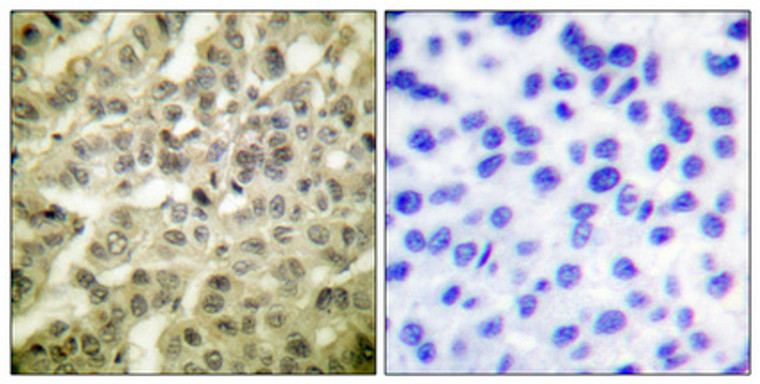
  • Immunohistochemistry analysis of paraffin-embedded human breast cancer, using Akt (Phospho-Thr308) Antibody. The picture on the right is blocked with the Akt (Phospho-Thr308) peptide.
  • Immunofluorescence analysis of rat-kidney tissue. 1, Akt (phospho Thr308) Polyclonal Antibody (red) was diluted at 1:200 (4°C, overnight). 2, Cy3 labled Secondary antibody was diluted at 1:300 (room temperature, 50min).3, Picture B: DAPI (blue) 10min. Picture A:Target. Picture B: DAPI. Picture C: merge of A+B
  • Immunohistochemical analysis of paraffin-embedded Human-uterus-cancer tissue. 1, Akt (phospho Thr308) Polyclonal Antibody was diluted at 1:200 (4°C, overnight). 2, Sodium citrate pH 6.0 was used for antibody retrieval (>98°C, 20min). 3, Secondary antibody was diluted at 1:200 (room tempeRature, 30min). Negative control was used by secondary antibody only.
  • Immunofluorescence analysis of rat-kidney tissue. 1, Akt (phospho Thr308) Polyclonal Antibody (red) was diluted at 1:200 (4°C, overnight). 2, Cy3 labled Secondary antibody was diluted at 1:300 (room temperature, 50min).3, Picture B: DAPI (blue) 10min. Picture A:Target. Picture B: DAPI. Picture C: merge of A+B
  • Immunofluorescence analysis of human-stomach tissue. 1, Akt (phospho Thr308) Polyclonal Antibody (red) was diluted at 1:200 (4°C, overnight). 2, Cy3 labled Secondary antibody was diluted at 1:300 (room temperature, 50min).3, Picture B: DAPI (blue) 10min. Picture A:Target. Picture B: DAPI. Picture C: merge of A+B
  • Immunofluorescence analysis of human-stomach tissue. 1, Akt (phospho Thr308) Polyclonal Antibody (red) was diluted at 1:200 (4°C, overnight). 2, Cy3 labled Secondary antibody was diluted at 1:300 (room temperature, 50min).3, Picture B: DAPI (blue) 10min. Picture A:Target. Picture B: DAPI. Picture C: merge of A+B
  • Western blot analysis of various cells using Phospho-Akt (T308) Polyclonal Antibody diluted at 1:500
  • Immunohistochemical analysis of paraffin-embedded Human-lung-cancer tissue. 1, Akt (phospho Thr308) Polyclonal Antibody was diluted at 1:200 (4°C, overnight). 2, Sodium citrate pH 6.0 was used for antibody retrieval (>98°C, 20min). 3, Secondary antibody was diluted at 1:200 (room tempeRature, 30min). Negative control was used by secondary antibody only.
  • Immunohistochemical analysis of paraffin-embedded Human breast cancer. Antibody was diluted at 1:100 (4°C overnight). High-pressure and temperature Tris-EDTA, pH8.0 was used for antigen retrieval. Negetive contrl (right) obtaned from antibody was pre-absorbed by immunogen peptide.
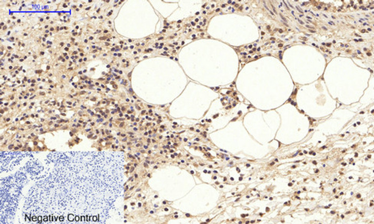
  • Immunohistochemical analysis of paraffin-embedded Human-Appendix tissue. 1, Akt (phospho Thr308) Polyclonal Antibody was diluted at 1:200 (4°C, overnight). 2, Sodium citrate pH 6.0 was used for antibody retrieval (>98°C, 20min). 3, Secondary antibody was diluted at 1:200 (room tempeRature, 30min). Negative control was used by secondary antibody only.
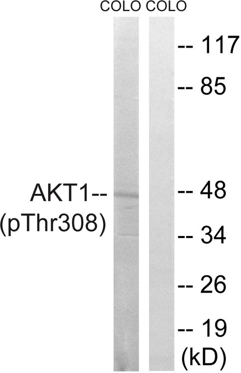
  • Western blot analysis of Akt (Phospho-Thr308) Antibody. The lane on the right is blocked with the Akt (Phospho-Thr308) peptide.
  • Immunofluorescence analysis of human-lung tissue. 1, Akt (phospho Thr308) Polyclonal Antibody (red) was diluted at 1:200 (4°C, overnight). 2, Cy3 labled Secondary antibody was diluted at 1:300 (room temperature, 50min).3, Picture B: DAPI (blue) 10min. Picture A:Target. Picture B: DAPI. Picture C: merge of A+B

Anti-Phospho-Akt-Thr308 antibody (STJ90750)

SKU:
STJ90750

Current Stock:
Host: Rabbit
Applications: IF/WB/IHC/ELISA
Reactivity: Human/Mouse/Rat/D.melanogaster
Note: STRICTLY FOR FURTHER SCIENTIFIC RESEARCH USE ONLY (RUO). MUST NOT TO BE USED IN DIAGNOSTIC OR THERAPEUTIC APPLICATIONS.
Short Description: Rabbit polyclonal antibody anti-Phospho-RAC-alpha serine/threonine-protein kinase and RAC-beta serine/threonine-protein kinase and RAC-gamma serine/threonine-protein kinase-Thr308 is suitable for use in Immunofluorescence, Western Blot, Immunohistoch
Clonality: Polyclonal
Conjugation: Unconjugated
Isotype: IgG
Formulation: Liquid in PBS containing 50% Glycerol, 0.5% BSA and 0.02% Sodium Azide.
Purification: The antibody was affinity-purified from rabbit antiserum by affinity-chromatography using epitope-specific immunogen.
Concentration: 1 mg/mL
Dilution Range: IF 1:50-200
WB 1:500-1:2000
IHC 1:100-1:300
ELISA 1:20000
Storage Instruction: Store at-20°C for up to 1 year from the date of receipt, and avoid repeat freeze-thaw cycles.
Gene Symbol: AKT3
AKT1
10000
207
208
AKT3_HUMAN
AKT1_HUMAN
AKT2_HUMAN
Specificity: Phospho-Akt (T308) Polyclonal Antibody detects endogenous levels of Akt protein only when phosphorylated at T308.
Immunogen: Synthesized phospho-peptide around the phosphorylation site of human Akt (phospho Thr308)
Post Translational Modifications O-GlcNAcylation at Thr-305 and Thr-312 inhibits activating phosphorylation at Thr-308 via disrupting the interaction between AKT1 and PDPK1. O-GlcNAcylation at Ser-473 also probably interferes with phosphorylation at this site. Phosphorylation on Thr-308, Ser-473 and Tyr-474 is required for full activity. Activated TNK2 phosphorylates it on Tyr-176 resulting in its binding to the anionic plasma membrane phospholipid PA. This phosphorylated form localizes to the cell membrane, where it is targeted by PDPK1 and PDPK2 for further phosphorylations on Thr-308 and Ser-473 leading to its activation. Ser-473 phosphorylation by mTORC2 favors Thr-308 phosphorylation by PDPK1. Phosphorylated at Thr-308 and Ser-473 by IKBKE and TBK1. Ser-473 phosphorylation is enhanced by interaction with AGAP2 isoform 2 (PIKE-A). Ser-473 phosphorylation is enhanced in focal cortical dysplasias with Taylor-type balloon cells. Ser-473 phosphorylation is enhanced by signaling through activated FLT3. Ser-473 is dephosphorylated by PHLPP. Dephosphorylated at Thr-308 and Ser-473 by PP2A phosphatase. The phosphorylated form of PPP2R5B is required for bridging AKT1 with PP2A phosphatase. Ser-473 is dephosphorylated by CPPED1, leading to termination of signaling. Ubiquitinated via 'Lys-48'-linked polyubiquitination by ZNRF1, leading to its degradation by the proteasome. Ubiquitinated.undergoes both 'Lys-48'- and 'Lys-63'-linked polyubiquitination. TRAF6-induced 'Lys-63'-linked AKT1 ubiquitination is critical for phosphorylation and activation. When ubiquitinated, it translocates to the plasma membrane, where it becomes phosphorylated. When fully phosphorylated and translocated into the nucleus, undergoes 'Lys-48'-polyubiquitination catalyzed by TTC3, leading to its degradation by the proteasome. Also ubiquitinated by TRIM13 leading to its proteasomal degradation. Phosphorylated, undergoes 'Lys-48'-linked polyubiquitination preferentially at Lys-284 catalyzed by MUL1, leading to its proteasomal degradation. Acetylated on Lys-14 and Lys-20 by the histone acetyltransferases EP300 and KAT2B. Acetylation results in reduced phosphorylation and inhibition of activity. Deacetylated at Lys-14 and Lys-20 by SIRT1. SIRT1-mediated deacetylation relieves the inhibition.
Function AKT1 is one of 3 closely related serine/threonine-protein kinases (AKT1, AKT2 and AKT3) called the AKT kinase, and which regulate many processes including metabolism, proliferation, cell survival, growth and angiogenesis. This is mediated through serine and/or threonine phosphorylation of a range of downstream substrates. Over 100 substrate candidates have been reported so far, but for most of them, no isoform specificity has been reported. AKT is responsible of the regulation of glucose uptake by mediating insulin-induced translocation of the SLC2A4/GLUT4 glucose transporter to the cell surface. Phosphorylation of PTPN1 at 'Ser-50' negatively modulates its phosphatase activity preventing dephosphorylation of the insulin receptor and the attenuation of insulin signaling. Phosphorylation of TBC1D4 triggers the binding of this effector to inhibitory 14-3-3 proteins, which is required for insulin-stimulated glucose transport. AKT regulates also the storage of glucose in the form of glycogen by phosphorylating GSK3A at 'Ser-21' and GSK3B at 'Ser-9', resulting in inhibition of its kinase activity. Phosphorylation of GSK3 isoforms by AKT is also thought to be one mechanism by which cell proliferation is driven. AKT regulates also cell survival via the phosphorylation of MAP3K5 (apoptosis signal-related kinase). Phosphorylation of 'Ser-83' decreases MAP3K5 kinase activity stimulated by oxidative stress and thereby prevents apoptosis. AKT mediates insulin-stimulated protein synthesis by phosphorylating TSC2 at 'Ser-939' and 'Thr-1462', thereby activating mTORC1 signaling and leading to both phosphorylation of 4E-BP1 and in activation of RPS6KB1. AKT is involved in the phosphorylation of members of the FOXO factors (Forkhead family of transcription factors), leading to binding of 14-3-3 proteins and cytoplasmic localization. In particular, FOXO1 is phosphorylated at 'Thr-24', 'Ser-256' and 'Ser-319'. FOXO3 and FOXO4 are phosphorylated on equivalent sites. AKT has an important role in the regulation of NF-kappa-B-dependent gene transcription and positively regulates the activity of CREB1 (cyclic AMP (cAMP)-response element binding protein). The phosphorylation of CREB1 induces the binding of accessory proteins that are necessary for the transcription of pro-survival genes such as BCL2 and MCL1. AKT phosphorylates 'Ser-454' on ATP citrate lyase (ACLY), thereby potentially regulating ACLY activity and fatty acid synthesis. Activates the 3B isoform of cyclic nucleotide phosphodiesterase (PDE3B) via phosphorylation of 'Ser-273', resulting in reduced cyclic AMP levels and inhibition of lipolysis. Phosphorylates PIKFYVE on 'Ser-318', which results in increased PI(3)P-5 activity. The Rho GTPase-activating protein DLC1 is another substrate and its phosphorylation is implicated in the regulation cell proliferation and cell growth. AKT plays a role as key modulator of the AKT-mTOR signaling pathway controlling the tempo of the process of newborn neurons integration during adult neurogenesis, including correct neuron positioning, dendritic development and synapse formation. Signals downstream of phosphatidylinositol 3-kinase (PI(3)K) to mediate the effects of various growth factors such as platelet-derived growth factor (PDGF), epidermal growth factor (EGF), insulin and insulin-like growth factor I (IGF-I). AKT mediates the antiapoptotic effects of IGF-I. Essential for the SPATA13-mediated regulation of cell migration and adhesion assembly and disassembly. May be involved in the regulation of the placental development. Phosphorylates STK4/MST1 at 'Thr-120' and 'Thr-387' leading to inhibition of its: kinase activity, nuclear translocation, autophosphorylation and ability to phosphorylate FOXO3. Phosphorylates STK3/MST2 at 'Thr-117' and 'Thr-384' leading to inhibition of its: cleavage, kinase activity, autophosphorylation at Thr-180, binding to RASSF1 and nuclear translocation. Phosphorylates SRPK2 and enhances its kinase activity towards SRSF2 and ACIN1 and promotes its nuclear translocation. Phosphorylates RAF1 at 'Ser-259' and negatively regulates its activity. Phosphorylation of BAD stimulates its pro-apoptotic activity. Phosphorylates KAT6A at 'Thr-369' and this phosphorylation inhibits the interaction of KAT6A with PML and negatively regulates its acetylation activity towards p53/TP53. Phosphorylates palladin (PALLD), modulating cytoskeletal organization and cell motility. Phosphorylates prohibitin (PHB), playing an important role in cell metabolism and proliferation. Phosphorylates CDKN1A, for which phosphorylation at 'Thr-145' induces its release from CDK2 and cytoplasmic relocalization. These recent findings indicate that the AKT1 isoform has a more specific role in cell motility and proliferation. Phosphorylates CLK2 thereby controlling cell survival to ionizing radiation. Phosphorylates PCK1 at 'Ser-90', reducing the binding affinity of PCK1 to oxaloacetate and changing PCK1 into an atypical protein kinase activity using GTP as donor.
Protein Name Rac-Alpha Serine/Threonine-Protein Kinase
Protein Kinase B
Pkb
Protein Kinase B Alpha
Pkb Alpha
Proto-Oncogene C-Akt
Rac-Pk-Alpha
Database Links Reactome: R-HSA-111447
Reactome: R-HSA-1257604
Reactome: R-HSA-1358803
Reactome: R-HSA-1445148
Reactome: R-HSA-1474151
Reactome: R-HSA-165159
Reactome: R-HSA-198323
Reactome: R-HSA-198693
Reactome: R-HSA-199418
Reactome: R-HSA-203615
Reactome: R-HSA-211163
Reactome: R-HSA-354192
Reactome: R-HSA-3769402
Reactome: R-HSA-389357
Reactome: R-HSA-389513
Reactome: R-HSA-392451
Reactome: R-HSA-450385
Reactome: R-HSA-450604
Reactome: R-HSA-5218920
Reactome: R-HSA-5628897
Reactome: R-HSA-5674400
Reactome: R-HSA-6785807
Reactome: R-HSA-6804757
Reactome: R-HSA-6804758
Reactome: R-HSA-6804759
Reactome: R-HSA-6811558
Reactome: R-HSA-69202
Reactome: R-HSA-69656
Reactome: R-HSA-8849469
Reactome: R-HSA-8876198
Reactome: R-HSA-8941332
Reactome: R-HSA-8948751
Reactome: R-HSA-9009391
Reactome: R-HSA-9604323
Reactome: R-HSA-9614399
Reactome: R-HSA-9634638
Cellular Localisation Cytoplasm
Nucleus
Cell Membrane
Nucleus After Activation By Integrin-Linked Protein Kinase 1 (Ilk1)
Nuclear Translocation Is Enhanced By Interaction With Tcl1a
Phosphorylation On Tyr-176 By Tnk2 Results In Its Localization To The Cell Membrane Where It Is Targeted For Further Phosphorylations On Thr-308 And Ser-473 Leading To Its Activation And The Activated Form Translocates To The Nucleus
Colocalizes With Wdfy2 In Intracellular Vesicles
Alternative Antibody Names Anti-Rac-Alpha Serine/Threonine-Protein Kinase antibody
Anti-Protein Kinase B antibody
Anti-Pkb antibody
Anti-Protein Kinase B Alpha antibody
Anti-Pkb Alpha antibody
Anti-Proto-Oncogene C-Akt antibody
Anti-Rac-Pk-Alpha antibody
Anti-AKT1 antibody
Anti-PKB antibody
Anti-RAC antibody

Information sourced from Uniprot.org

12 months for antibodies. 6 months for ELISA Kits. Please see website T&Cs for further guidance