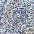
• Immunohistochemistry analysis of PAXIP1 in paraffin-embedded mouse spleen using PAXIP1 Rabbit polyclonal antibody (STJ11102906) at dilution of 1:100 (40x lens). Perform high pressure antigen retrieval with 10 mM citrate buffer pH 6. 0 before commencing with immunohistochemistry staining protocol.
  • Immunohistochemistry analysis of PAXIP1 in paraffin-embedded mouse brain using PAXIP1 Rabbit polyclonal antibody (STJ11102906) at dilution of 1:100 (40x lens). Perform high pressure antigen retrieval with 10 mM citrate buffer pH 6. 0 before commencing with immunohistochemistry staining protocol.
  • Immunohistochemistry analysis of PAXIP1 in paraffin-embedded human breast cancer using PAXIP1 Rabbit polyclonal antibody (STJ11102906) at dilution of 1:100 (40x lens). Perform high pressure antigen retrieval with 10 mM citrate buffer pH 6. 0 before commencing with immunohistochemistry staining protocol.
  • Immunohistochemistry analysis of PAXIP1 in paraffin-embedded human thyroid cancer using PAXIP1 Rabbit polyclonal antibody (STJ11102906) at dilution of 1:100 (40x lens). Perform high pressure antigen retrieval with 10 mM citrate buffer pH 6. 0 before commencing with immunohistochemistry staining protocol.
  • Immunohistochemistry analysis of PAXIP1 in paraffin-embedded rat spleen using PAXIP1 Rabbit polyclonal antibody (STJ11102906) at dilution of 1:100 (40x lens). Perform high pressure antigen retrieval with 10 mM citrate buffer pH 6. 0 before commencing with immunohistochemistry staining protocol.
  • Immunohistochemistry analysis of PAXIP1 in paraffin-embedded rat ovary using PAXIP1 Rabbit polyclonal antibody (STJ11102906) at dilution of 1:100 (40x lens). Perform high pressure antigen retrieval with 10 mM citrate buffer pH 6. 0 before commencing with immunohistochemistry staining protocol.
  • Western blot analysis of various lysates using PAXIP1 Rabbit polyclonal antibody (STJ11102906) at 1:500 dilution. Secondary antibody: HRP Goat Anti-Rabbit IgG (H+L) (STJS000856) at 1:10000 dilution. Lysates/proteins: 25 Mu g per lane. Blocking buffer: 3% nonfat dry milk in TBST. Detection: ECL Enhanced Kit. Exposure time: 180s.

Anti-PAXIP1 antibody (301-394) (STJ11102906)

SKU:
STJ11102906

Shipping:
Free Shipping
Current Stock:
Host: Rabbit
Applications: WB/IHC
Reactivity: Human/Mouse/Rat
Note: STRICTLY FOR FURTHER SCIENTIFIC RESEARCH USE ONLY (RUO). MUST NOT TO BE USED IN DIAGNOSTIC OR THERAPEUTIC APPLICATIONS.
Short Description: Rabbit polyclonal antibody anti-PAXIP1 (301-394) is suitable for use in Western Blot and Immunohistochemistry research applications.
Clonality: Polyclonal
Conjugation: Unconjugated
Isotype: IgG
Formulation: PBS with 0.01% Thimerosal, 50% Glycerol, pH7.3.
Purification: Affinity purification
Dilution Range: WB 1:100-1:500
IHC-P 1:50-1:200
Storage Instruction: Store at-20°C for up to 1 year from the date of receipt, and avoid repeat freeze-thaw cycles.
Gene Symbol: PAXIP1
Gene ID: 22976
Uniprot ID: PAXI1_HUMAN
Immunogen Region: 301-394
Immunogen: Recombinant fusion protein containing a sequence corresponding to amino acids 301-394 of human PAXIP1 (NP_031375.3).
Immunogen Sequence: GNILPPEVRGNLMAAGQNLQ SSERSEMIATWSPAVRTLRN ITNNADIQQMNRPSNVAHIL QTLSAPTKNLEQQVNHSQQG HTNANAVLFSQVKV
Function Involved in DNA damage response and in transcriptional regulation through histone methyltransferase (HMT) complexes. Plays a role in early development. In DNA damage response is required for cell survival after ionizing radiation. In vitro shown to be involved in the homologous recombination mechanism for the repair of double-strand breaks (DSBs). Its localization to DNA damage foci requires RNF8 and UBE2N. Recruits TP53BP1 to DNA damage foci and, at least in particular repair processes, effective DNA damage response appears to require the association with TP53BP1 phosphorylated by ATM at 'Ser-25'. Together with TP53BP1 regulates ATM association. Proposed to recruit PAGR1 to sites of DNA damage and the PAGR1:PAXIP1 complex is required for cell survival in response to DNA damage.the function is probably independent of MLL-containing histone methyltransferase (HMT) complexes. However, this function has been questioned. Promotes ubiquitination of PCNA following UV irradiation and may regulate recruitment of polymerase eta and RAD51 to chromatin after DNA damage. Proposed to be involved in transcriptional regulation by linking MLL-containing histone methyltransferase (HMT) complexes to gene promoters by interacting with promoter-bound transcription factors such as PAX2. Associates with gene promoters that are known to be regulated by KMT2D/MLL2. During immunoglobulin class switching in activated B-cells is involved in trimethylation of histone H3 at 'Lys-4' and in transcription initiation of downstream switch regions at the immunoglobulin heavy-chain (Igh) locus.this function appears to involve the recruitment of MLL-containing HMT complexes. Conflictingly, its function in transcriptional regulation during immunoglobulin class switching is reported to be independent of the MLL2/MLL3 complex.
Protein Name Pax-Interacting Protein 1
Pax Transactivation Activation Domain-Interacting Protein
Database Links Reactome: R-HSA-5617472
Reactome: R-HSA-5693571
Reactome: R-HSA-9772755
Cellular Localisation Nucleus Matrix
Chromosome
Localizes To Dna Damage Foci Upon Ionizing Radiation
Alternative Antibody Names Anti-Pax-Interacting Protein 1 antibody
Anti-Pax Transactivation Activation Domain-Interacting Protein antibody
Anti-PAXIP1 antibody
Anti-PAXIP1L antibody
Anti-PTIP antibody
Anti-CAGF28 antibody

Information sourced from Uniprot.org

12 months for antibodies. 6 months for ELISA Kits. Please see website T&Cs for further guidance