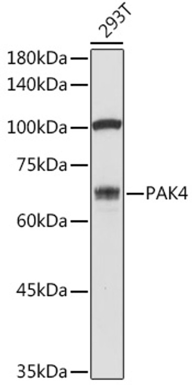
| Host: |
Rabbit |
| Applications: |
WB |
| Reactivity: |
Human |
| Note: |
STRICTLY FOR FURTHER SCIENTIFIC RESEARCH USE ONLY (RUO). MUST NOT TO BE USED IN DIAGNOSTIC OR THERAPEUTIC APPLICATIONS. |
| Short Description: |
Rabbit polyclonal antibody anti-PAK4 (120-591) is suitable for use in Western Blot research applications. |
| Clonality: |
Polyclonal |
| Conjugation: |
Unconjugated |
| Isotype: |
IgG |
| Formulation: |
PBS with 0.01% Thimerosal, 50% Glycerol, pH7.3. |
| Purification: |
Affinity purification |
| Dilution Range: |
WB 1:500-1:1000 |
| Storage Instruction: |
Store at-20°C for up to 1 year from the date of receipt, and avoid repeat freeze-thaw cycles. |
| Gene Symbol: |
PAK4 |
| Gene ID: |
10298 |
| Uniprot ID: |
PAK4_HUMAN |
| Immunogen Region: |
120-591 |
| Immunogen: |
Recombinant fusion protein containing a sequence corresponding to amino acids 120-591 of human PAK4 (NP_005875.1). |
| Immunogen Sequence: |
EPATTARGGPGKAGSRGRFA GHSEAGGGSGDRRRAGPEKR PKSSREGSGGPQESSRDKRP LSGPDVGTPQPAGLASGAKL AAGRPFNTYPRADTDHPSRG AQGEPHDVAPNGPSAGGLAI PQSSSSSSRPPTRARGAPSP GVLGPHASEPQLAPPACTPA A |
| Tissue Specificity | Highest expression in prostate, testis and colon. |
| Post Translational Modifications | Autophosphorylated on serine residues when activated by CDC42/p21 (Ref.32). Phosphorylated on tyrosine residues upon stimulation of FGFR2. Methylated by SETD6. Polyubiquitinated, leading to its proteasomal degradation. |
| Function | Serine/threonine protein kinase that plays a role in a variety of different signaling pathways including cytoskeleton regulation, cell migration, growth, proliferation or cell survival. Activation by various effectors including growth factor receptors or active CDC42 and RAC1 results in a conformational change and a subsequent autophosphorylation on several serine and/or threonine residues. Phosphorylates and inactivates the protein phosphatase SSH1, leading to increased inhibitory phosphorylation of the actin binding/depolymerizing factor cofilin. Decreased cofilin activity may lead to stabilization of actin filaments. Phosphorylates LIMK1, a kinase that also inhibits the activity of cofilin. Phosphorylates integrin beta5/ITGB5 and thus regulates cell motility. Phosphorylates ARHGEF2 and activates the downstream target RHOA that plays a role in the regulation of assembly of focal adhesions and actin stress fibers. Stimulates cell survival by phosphorylating the BCL2 antagonist of cell death BAD. Alternatively, inhibits apoptosis by preventing caspase-8 binding to death domain receptors in a kinase independent manner. Plays a role in cell-cycle progression by controlling levels of the cell-cycle regulatory protein CDKN1A and by phosphorylating RAN. |
| Protein Name | Serine/Threonine-Protein Kinase Pak 4P21-Activated Kinase 4Pak-4 |
| Database Links | Reactome: R-HSA-428540Reactome: R-HSA-9013148Reactome: R-HSA-9013149Reactome: R-HSA-9013404Reactome: R-HSA-9013406Reactome: R-HSA-9013407Reactome: R-HSA-9013408Reactome: R-HSA-9013409Reactome: R-HSA-9013420Reactome: R-HSA-9013423Reactome: R-HSA-9013424 |
| Cellular Localisation | CytoplasmSeems To Shuttle Between Cytoplasmic Compartments Depending On The Activating EffectorFor ExampleCan Be Found On The Cell Periphery After Activation Of Growth-Factor Or Integrin-Mediated Signaling Pathways |
| Alternative Antibody Names | Anti-Serine/Threonine-Protein Kinase Pak 4 antibodyAnti-P21-Activated Kinase 4 antibodyAnti-Pak-4 antibodyAnti-PAK4 antibodyAnti-KIAA1142 antibody |
Information sourced from Uniprot.org
12 months for antibodies. 6 months for ELISA Kits. Please see website T&Cs for further guidance


