| Host: |
Rabbit |
| Applications: |
WB/IF |
| Reactivity: |
Human/Mouse/Rat |
| Note: |
STRICTLY FOR FURTHER SCIENTIFIC RESEARCH USE ONLY (RUO). MUST NOT TO BE USED IN DIAGNOSTIC OR THERAPEUTIC APPLICATIONS. |
| Short Description: |
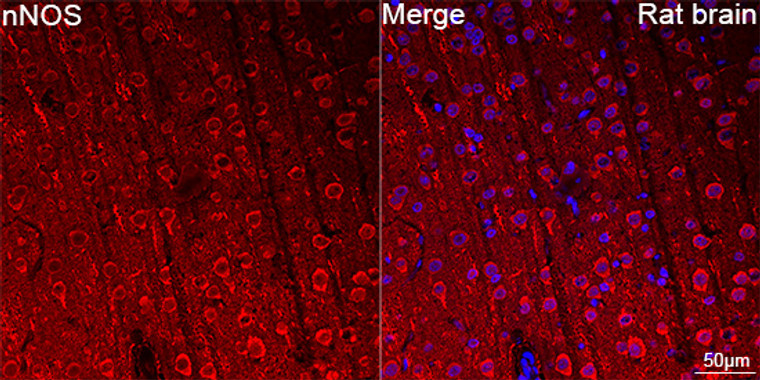
Rabbit monoclonal antibody anti-nNOS (1335-1434) is suitable for use in Western Blot and Immunofluorescence research applications. |
| Clonality: |
Monoclonal |
| Clone ID: |
S9MR |
| Conjugation: |
Unconjugated |
| Isotype: |
IgG |
| Formulation: |
PBS with 0.02% Sodium Azide, 0.05% BSA, 50% Glycerol, pH7.3. |
| Purification: |
Affinity purification |
| Dilution Range: |
WB 1:500-1:1000IF/ICC 1:50-1:200 |
| Storage Instruction: |
Store at-20°C for up to 1 year from the date of receipt, and avoid repeat freeze-thaw cycles. |
| Gene Symbol: |
NOS1 |
| Gene ID: |
4842 |
| Uniprot ID: |
NOS1_HUMAN |
| Immunogen Region: |
1335-1434 |
| Immunogen: |
A synthetic peptide corresponding to a sequence within amino acids 1335-1434 of human nNOS (P29475). |
| Immunogen Sequence: |
QLAESVYRALKEQGGHIYVC GDVTMAADVLKAIQRIMTQQ GKLSAEDAGVFISRMRDDNR YHEDIFGVTLRTYEVTNRLR SESIAFIEESKKDTDEVFSS |
| Tissue Specificity | Isoform 1 is ubiquitously expressed: detected in skeletal muscle and brain, also in testis, lung and kidney, and at low levels in heart, adrenal gland and retina. Not detected in the platelets. Isoform 3 is expressed only in testis. Isoform 4 is detected in testis, skeletal muscle, lung, and kidney, at low levels in the brain, but not in the heart and adrenal gland. |
| Post Translational Modifications | Ubiquitinated.mediated by STUB1/CHIP in the presence of Hsp70 and Hsp40 (in vitro). |
| Function | Produces nitric oxide (NO) which is a messenger molecule with diverse functions throughout the body. In the brain and peripheral nervous system, NO displays many properties of a neurotransmitter. Probably has nitrosylase activity and mediates cysteine S-nitrosylation of cytoplasmic target proteins such SRR. |
| Protein Name | Nitric Oxide Synthase 1Constitutive NosNc-NosNos Type INeuronal NosN-NosNnosNitric Oxide Synthase - BrainBnosPeptidyl-Cysteine S-Nitrosylase Nos1 |
| Database Links | Reactome: R-HSA-1222556Reactome: R-HSA-392154Reactome: R-HSA-5578775 |
| Cellular Localisation | Cell MembraneSarcolemmaPeripheral Membrane ProteinCell ProjectionDendritic SpineIn Skeletal MuscleIt Is Localized Beneath The Sarcolemma Of Fast-Twitch Muscle Fiber By Associating With The Dystrophin Glycoprotein ComplexIn NeuronsEnriched In Dendritic Spines |
| Alternative Antibody Names | Anti-Nitric Oxide Synthase 1 antibodyAnti-Constitutive Nos antibodyAnti-Nc-Nos antibodyAnti-Nos Type I antibodyAnti-Neuronal Nos antibodyAnti-N-Nos antibodyAnti-Nnos antibodyAnti-Nitric Oxide Synthase - Brain antibodyAnti-Bnos antibodyAnti-Peptidyl-Cysteine S-Nitrosylase Nos1 antibodyAnti-NOS1 antibody |
Information sourced from Uniprot.org
12 months for antibodies. 6 months for ELISA Kits. Please see website T&Cs for further guidance


