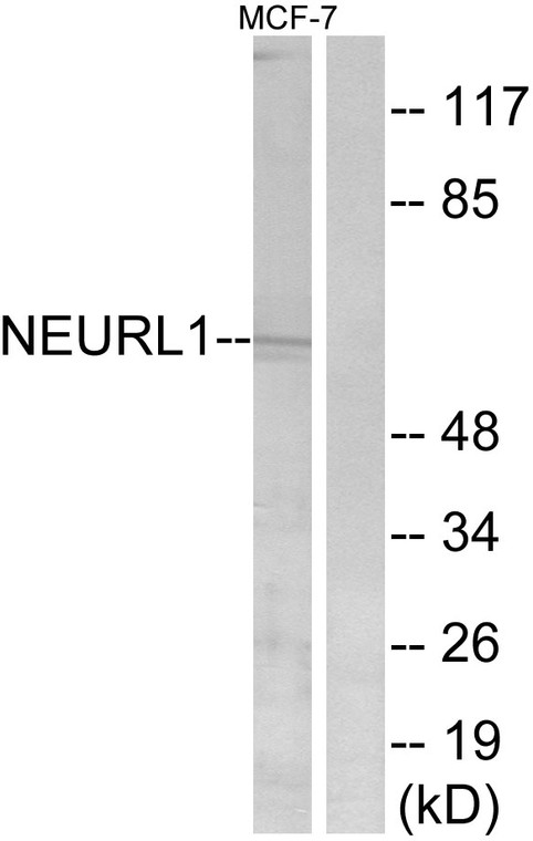
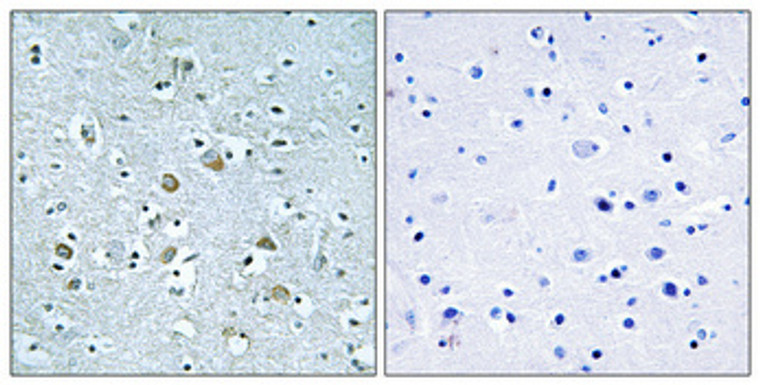
| Host: |
Rabbit |
| Applications: |
WB/IHC/IF/ELISA |
| Reactivity: |
Human/Rat/Mouse |
| Note: |
STRICTLY FOR FURTHER SCIENTIFIC RESEARCH USE ONLY (RUO). MUST NOT TO BE USED IN DIAGNOSTIC OR THERAPEUTIC APPLICATIONS. |
| Short Description: |
Rabbit polyclonal antibody anti-E3 ubiquitin-protein ligase NEURL1 (219-268) is suitable for use in Western Blot, Immunohistochemistry, Immunofluorescence and ELISA research applications. |
| Clonality: |
Polyclonal |
| Conjugation: |
Unconjugated |
| Isotype: |
IgG |
| Formulation: |
Liquid in PBS containing 50% Glycerol, 0.5% BSA and 0.02% Sodium Azide. |
| Purification: |
The antibody was affinity-purified from rabbit antiserum by affinity-chromatography using epitope-specific immunogen. |
| Concentration: |
1 mg/mL |
| Dilution Range: |
WB 1:500-1:2000IHC 1:100-1:300ELISA 1:40000IF 1:50-200 |
| Storage Instruction: |
Store at-20°C for up to 1 year from the date of receipt, and avoid repeat freeze-thaw cycles. |
| Gene Symbol: |
NEURL1 |
| Gene ID: |
9148 |
| Uniprot ID: |
NEUL1_HUMAN |
| Immunogen Region: |
219-268 |
| Specificity: |
Neuralized-1 Polyclonal Antibody detects endogenous levels of Neuralized-1 protein. |
| Immunogen: |
The antiserum was produced against synthesized peptide derived from human NEURL1. AA range:219-268 |
| Tissue Specificity | Expressed in brain, testis, pituitary gland, pancreas and bone marrow. Also poorly expressed in malignant astrocytomas and several neuroectodermal tumor cell lines. Weakly expressed in medulloblastoma (MB) compared with normal cerebellar tissues. |
| Post Translational Modifications | Myristoylation is a determinant of membrane targeting. |
| Function | Plays a role in hippocampal-dependent synaptic plasticity, learning and memory. Involved in the formation of spines and functional synaptic contacts by modulating the translational activity of the cytoplasmic polyadenylation element-binding protein CPEB3. Promotes ubiquitination of CPEB3, and hence induces CPEB3-dependent mRNA translation activation of glutamate receptor GRIA1 and GRIA2. Can function as an E3 ubiquitin-protein ligase to activate monoubiquitination of JAG1 (in vitro), thereby regulating the Notch pathway. Acts as a tumor suppressor.inhibits malignant cell transformation of medulloblastoma (MB) cells by inhibiting the Notch signaling pathway. |
| Protein Name | E3 Ubiquitin-Protein Ligase Neurl1Neuralized-Like Protein 1aH-NeuH-Neuralized 1Ring Finger Protein 67Ring-Type E3 Ubiquitin Transferase Neurl1 |
| Database Links | Reactome: R-HSA-2122948Reactome: R-HSA-2644606Reactome: R-HSA-2691232Reactome: R-HSA-2894862Reactome: R-HSA-2979096Reactome: R-HSA-9013507 |
| Cellular Localisation | CytoplasmPerinuclear RegionCell MembranePeripheral Membrane ProteinPerikaryonCell ProjectionDendritePostsynaptic DensityLocalized In The Cell Bodies Of The Pyramidal Neurons And Distributed Along Their Apical DendritesColocalized With Psd95 In Postsynaptic SitesColocalized With Cpeb3 At Apical Dendrites Of Ca1 NeuronsColocalized With Jag1 At The Cell Surface |
| Alternative Antibody Names | Anti-E3 Ubiquitin-Protein Ligase Neurl1 antibodyAnti-Neuralized-Like Protein 1a antibodyAnti-H-Neu antibodyAnti-H-Neuralized 1 antibodyAnti-Ring Finger Protein 67 antibodyAnti-Ring-Type E3 Ubiquitin Transferase Neurl1 antibodyAnti-NEURL1 antibodyAnti-NEURL antibodyAnti-NEURL1A antibodyAnti-RNF67 antibody |
Information sourced from Uniprot.org
12 months for antibodies. 6 months for ELISA Kits. Please see website T&Cs for further guidance


