| Host: |
Rabbit |
| Applications: |
WB/IF/IP |
| Reactivity: |
Human/Mouse/Rat |
| Note: |
STRICTLY FOR FURTHER SCIENTIFIC RESEARCH USE ONLY (RUO). MUST NOT TO BE USED IN DIAGNOSTIC OR THERAPEUTIC APPLICATIONS. |
| Short Description: |
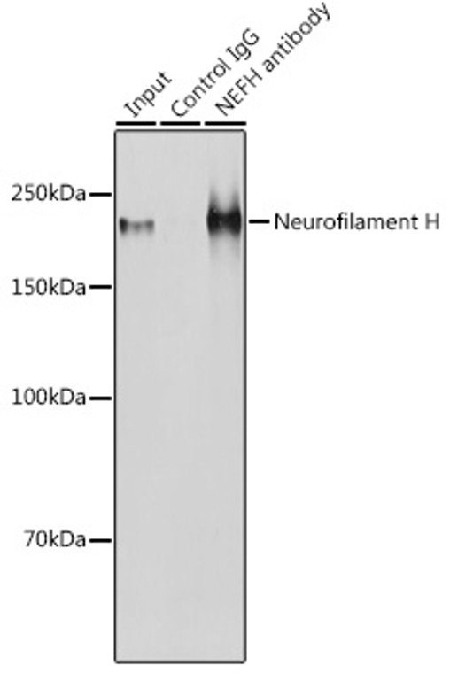
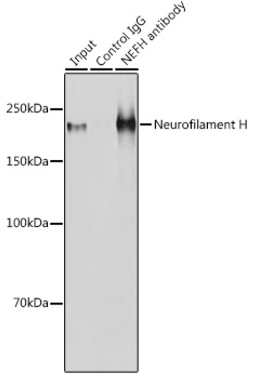
Rabbit polyclonal antibody anti-NEFH (873-1020) is suitable for use in Western Blot, Immunofluorescence and Immunoprecipitation research applications. |
| Clonality: |
Polyclonal |
| Conjugation: |
Unconjugated |
| Isotype: |
IgG |
| Formulation: |
PBS with 0.01% Thimerosal, 50% Glycerol, pH7.3. |
| Purification: |
Affinity purification |
| Dilution Range: |
WB 1:500-1:1000IF/ICC 1:50-1:200IP 1:500-1:1000 |
| Storage Instruction: |
Store at-20°C for up to 1 year from the date of receipt, and avoid repeat freeze-thaw cycles. |
| Gene Symbol: |
NEFH |
| Gene ID: |
4744 |
| Uniprot ID: |
NFH_HUMAN |
| Immunogen Region: |
873-1020 |
| Immunogen: |
Recombinant fusion protein containing a sequence corresponding to amino acids 873-1020 of human Neurofilament H (NP_066554.2). |
| Immunogen Sequence: |
KVEEKKEPAVEKPKESKVEA KKEEAEDKKKVPTPEKEAPA KVEVKEDAKPKEKTEVAKKE PDDAKAKEPSKPAEKKEAAP EKKDTKEEKAKKPEEKPKTE AKAKEDDKTLSKEPSKPKAE KAEKSSSTDQKDSKPPEKAT EDKAAKGK |
| Post Translational Modifications | There are a number of repeats of the tripeptide K-S-P, NFH is phosphorylated on a number of the serines in this motif. It is thought that phosphorylation of NFH results in the formation of interfilament cross bridges that are important in the maintenance of axonal caliber. Phosphorylation seems to play a major role in the functioning of the larger neurofilament polypeptides (NF-M and NF-H), the levels of phosphorylation being altered developmentally and coincidentally with a change in the neurofilament function. Phosphorylated in the head and rod regions by the PKC kinase PKN1, leading to the inhibition of polymerization. |
| Function | Neurofilaments usually contain three intermediate filament proteins: NEFL, NEFM, and NEFH which are involved in the maintenance of neuronal caliber. NEFH has an important function in mature axons that is not subserved by the two smaller NF proteins. May additionally cooperate with the neuronal intermediate filament proteins PRPH and INA to form neuronal filamentous networks. |
| Protein Name | Neurofilament Heavy PolypeptideNf-H200 Kda Neurofilament ProteinNeurofilament Triplet H Protein |
| Cellular Localisation | CytoplasmCytoskeletonCell ProjectionAxon |
| Alternative Antibody Names | Anti-Neurofilament Heavy Polypeptide antibodyAnti-Nf-H antibodyAnti-200 Kda Neurofilament Protein antibodyAnti-Neurofilament Triplet H Protein antibodyAnti-NEFH antibodyAnti-KIAA0845 antibodyAnti-NFH antibody |
Information sourced from Uniprot.org
12 months for antibodies. 6 months for ELISA Kits. Please see website T&Cs for further guidance


