| Host: |
Rabbit |
| Applications: |
WB |
| Reactivity: |
Human/Mouse/Rat |
| Note: |
STRICTLY FOR FURTHER SCIENTIFIC RESEARCH USE ONLY (RUO). MUST NOT TO BE USED IN DIAGNOSTIC OR THERAPEUTIC APPLICATIONS. |
| Short Description: |
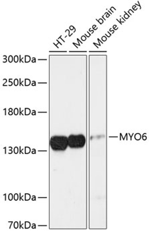
Rabbit polyclonal antibody anti-MYO6 (1016-1285) is suitable for use in Western Blot research applications. |
| Clonality: |
Polyclonal |
| Conjugation: |
Unconjugated |
| Isotype: |
IgG |
| Formulation: |
PBS with 0.01% Thimerosal, 50% Glycerol, pH7.3. |
| Purification: |
Affinity purification |
| Dilution Range: |
WB 1:500-1:2000 |
| Storage Instruction: |
Store at-20°C for up to 1 year from the date of receipt, and avoid repeat freeze-thaw cycles. |
| Gene Symbol: |
MYO6 |
| Gene ID: |
4646 |
| Uniprot ID: |
MYO6_HUMAN |
| Immunogen Region: |
1016-1285 |
| Immunogen: |
Recombinant fusion protein containing a sequence corresponding to amino acids 1016-1285 of human MYO6 (NP_004990.3). |
| Immunogen Sequence: |
IAQSEAELISDEAQADLALR RNDGTRPKMTPEQMAKEMSE FLSRGPAVLATKAAAGTKKY DLSKWKYAELRDTINTSCDI ELLAACREEFHRRLKVYHAW KSKNKKRNTETEQRAPKSVT DYDFAPFLNNSPQQNPAAQI PARQREIEMNRQQRFFRIPF IRPADQYKDPQSKKKGWWYA HFDGPWIARQMELHPDKPPI LLVAGKDDMEMCELNLEETG LTRKRGAEILPRQFEEIWE |
| Tissue Specificity | Expressed in most tissues examined including heart, brain, placenta, pancreas, spleen, thymus, prostate, testis, ovary, small intestine and colon. Highest levels in brain, pancreas, testis and small intestine. Also expressed in fetal brain and cochlea. Isoform 1 and isoform 2, containing the small insert, and isoform 4, containing neither insert, are expressed in unpolarized epithelial cells. |
| Post Translational Modifications | Phosphorylation in the motor domain, induced by EGF, results in translocation of MYO6 from the cell surface to membrane ruffles and affects F-actin dynamics. Phosphorylated in vitro by p21-activated kinase (PAK). |
| Function | Myosins are actin-based motor molecules with ATPase activity. Unconventional myosins serve in intracellular movements. Myosin 6 is a reverse-direction motor protein that moves towards the minus-end of actin filaments. Has slow rate of actin-activated ADP release due to weak ATP binding. Functions in a variety of intracellular processes such as vesicular membrane trafficking and cell migration. Required for the structural integrity of the Golgi apparatus via the p53-dependent pro-survival pathway. Appears to be involved in a very early step of clathrin-mediated endocytosis in polarized epithelial cells. Together with TOM1, mediates delivery of endocytic cargo to autophagosomes thereby promoting autophagosome maturation and driving fusion with lysosomes. Links TOM1 with autophagy receptors, such as TAX1BP1.CALCOCO2/NDP52 and OPTN. May act as a regulator of F-actin dynamics. As part of the DISP complex, may regulate the association of septins with actin and thereby regulate the actin cytoskeleton. May play a role in transporting DAB2 from the plasma membrane to specific cellular targets. May play a role in the extension and network organization of neurites. Required for structural integrity of inner ear hair cells. Modulates RNA polymerase II-dependent transcription. |
| Protein Name | Unconventional Myosin-ViUnconventional Myosin-6 |
| Database Links | Reactome: R-HSA-190873Reactome: R-HSA-399719Reactome: R-HSA-9013418Reactome: R-HSA-9013420Reactome: R-HSA-9013422 |
| Cellular Localisation | Golgi ApparatusTrans-Golgi Network MembranePeripheral Membrane ProteinNucleusCytoplasmPerinuclear RegionMembraneClathrin-Coated PitCytoplasmic VesicleClathrin-Coated VesicleCell ProjectionFilopodiumRuffle MembraneMicrovillusCytosolAutophagosomeEndosomeAlso Present In Endocyctic VesiclesTranslocates From Membrane RufflesEndocytic Vesicles And Cytoplasm To Golgi ApparatusPerinuclear Membrane And Nucleus Through Induction By P53 And P53-Induced Dna DamageRecruited Into Membrane Ruffles From Cell Surface By Egf-StimulationColocalizes With Dab2 In Clathrin-Coated Pits/VesiclesColocalizes With Optn At The Golgi Complex And In Vesicular Structures Close To The Plasma MembraneRecruited To Endosomes By Tom1 And Tom1l2Isoform 3: Cytoplasmic VesicleClathrin-Coated Vesicle MembraneIsoform 4: Cytoplasmic Vesicle |
| Alternative Antibody Names | Anti-Unconventional Myosin-Vi antibodyAnti-Unconventional Myosin-6 antibodyAnti-MYO6 antibodyAnti-KIAA0389 antibody |
Information sourced from Uniprot.org
12 months for antibodies. 6 months for ELISA Kits. Please see website T&Cs for further guidance


