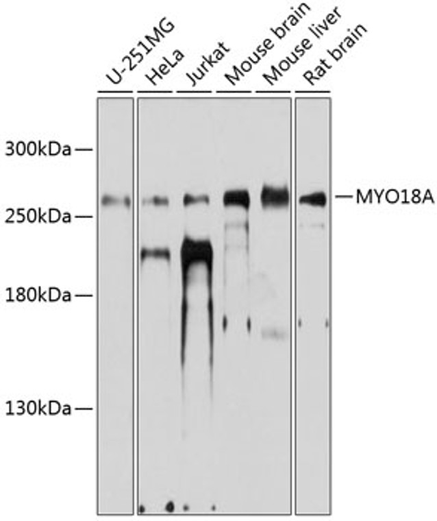
| Host: |
Rabbit |
| Applications: |
WB |
| Reactivity: |
Human/Mouse/Rat |
| Note: |
STRICTLY FOR FURTHER SCIENTIFIC RESEARCH USE ONLY (RUO). MUST NOT TO BE USED IN DIAGNOSTIC OR THERAPEUTIC APPLICATIONS. |
| Short Description: |
Rabbit polyclonal antibody anti-MYO18A (1970-2054) is suitable for use in Western Blot research applications. |
| Clonality: |
Polyclonal |
| Conjugation: |
Unconjugated |
| Isotype: |
IgG |
| Formulation: |
PBS with 0.01% Thimerosal, 50% Glycerol, pH7.3. |
| Purification: |
Affinity purification |
| Dilution Range: |
WB 1:1000-1:2000 |
| Storage Instruction: |
Store at-20°C for up to 1 year from the date of receipt, and avoid repeat freeze-thaw cycles. |
| Gene Symbol: |
MYO18A |
| Gene ID: |
399687 |
| Uniprot ID: |
MY18A_HUMAN |
| Immunogen Region: |
1970-2054 |
| Immunogen: |
Recombinant fusion protein containing a sequence corresponding to amino acids 1970-2054 of human MYO18A (NP_510880.2). |
| Immunogen Sequence: |
SDVDSELEDRVDGVKSWLSK NKGPSKAASDDGSLKSSSPT SYWKSLAPDRSDDEHDPLDN TSRPRYSHSYLSDSDTEAKL TETNA |
| Function | May link Golgi membranes to the cytoskeleton and participate in the tensile force required for vesicle budding from the Golgi. Thereby, may play a role in Golgi membrane trafficking and could indirectly give its flattened shape to the Golgi apparatus. Alternatively, in concert with LURAP1 and CDC42BPA/CDC42BPB, has been involved in modulating lamellar actomyosin retrograde flow that is crucial to cell protrusion and migration. May be involved in the maintenance of the stromal cell architectures required for cell to cell contact. Regulates trafficking, expression, and activation of innate immune receptors on macrophages. Plays a role to suppress inflammatory responsiveness of macrophages via a mechanism that modulates CD14 trafficking. Acts as a receptor of surfactant-associated protein A (SFTPA1/SP-A) and plays an important role in internalization and clearance of SFTPA1-opsonized S.aureus by alveolar macrophages. Strongly enhances natural killer cell cytotoxicity. |
| Protein Name | Unconventional Myosin-XviiiaMolecule Associated With Jak3 N-TerminusMajnMyosin Containing A Pdz DomainSurfactant Protein Receptor Sp-R210Sp-R210 |
| Database Links | Reactome: R-HSA-1839117Reactome: R-HSA-5655302Reactome: R-HSA-9703465 |
| Cellular Localisation | Golgi ApparatusTrans-Golgi NetworkGolgi OutpostCytoplasmCytoskeletonMicrotubule Organizing CenterRecruited To The Golgi Apparatus By Golph3Localizes To The Postsynaptic Golgi Apparatus RegionAlso Named Golgi OutpostWhich Shapes Dendrite Morphology By Functioning As Sites Of Acentrosomal Microtubule NucleationIsoform 1: Endoplasmic Reticulum-Golgi Intermediate CompartmentColocalizes With ActinIsoform 2: CytoplasmLacks The Pdz DomainDiffusely Localized In The CytoplasmIsoform 5: Cell Surface |
| Alternative Antibody Names | Anti-Unconventional Myosin-Xviiia antibodyAnti-Molecule Associated With Jak3 N-Terminus antibodyAnti-Majn antibodyAnti-Myosin Containing A Pdz Domain antibodyAnti-Surfactant Protein Receptor Sp-R210 antibodyAnti-Sp-R210 antibodyAnti-MYO18A antibodyAnti-CD245 antibodyAnti-KIAA0216 antibodyAnti-MYSPDZ antibodyAnti-TIAF1 antibody |
Information sourced from Uniprot.org
12 months for antibodies. 6 months for ELISA Kits. Please see website T&Cs for further guidance


