• Western blot analysis of lysate from COLO cells, using MyD88 antibody.
  • Western blot analysis of mouse-liver mouse-heart mouse-lung Cell Lysate, antibody was diluted at 1:500. Secondary antibody was diluted at 1:20000
  • Immunohistochemistry analysis of MyD88 antibody in paraffin-embedded human brain tissue.
  • Western blot analysis of various cells using MyD88 Polyclonal Antibody diluted at 1:2000
  • Western blot analysis of COLO205 cells using MyD88 Polyclonal Antibody diluted at 1:2000
  • Immunohistochemical analysis of paraffin-embedded Rat-spleen tissue. 1, MyD88 Polyclonal Antibody was diluted at 1:200 (4°C, overnight). 2, Sodium citrate pH 6.0 was used for antibody retrieval (>98°C, 20min). 3, Secondary antibody was diluted at 1:200 (room tempeRature, 30min). Negative control was used by secondary antibody only.
  • Immunohistochemical analysis of paraffin-embedded Mouse-kidney tissue. 1, MyD88 Polyclonal Antibody was diluted at 1:200 (4°C, overnight). 2, Sodium citrate pH 6.0 was used for antibody retrieval (>98°C, 20min). 3, Secondary antibody was diluted at 1:200 (room tempeRature, 30min). Negative control was used by secondary antibody only.
  • Immunohistochemical analysis of paraffin-embedded Rat-kidney tissue. 1, MyD88 Polyclonal Antibody was diluted at 1:200 (4°C, overnight). 2, Sodium citrate pH 6.0 was used for antibody retrieval (>98°C, 20min). 3, Secondary antibody was diluted at 1:200 (room tempeRature, 30min). Negative control was used by secondary antibody only.
  • Immunohistochemical analysis of paraffin-embedded Rat-lung tissue. 1, MyD88 Polyclonal Antibody was diluted at 1:200 (4°C, overnight). 2, Sodium citrate pH 6.0 was used for antibody retrieval (>98°C, 20min). 3, Secondary antibody was diluted at 1:200 (room tempeRature, 30min). Negative control was used by secondary antibody only.
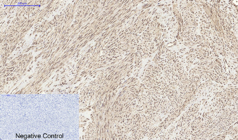
  • Immunohistochemical analysis of paraffin-embedded Human-uterus tissue. 1, MyD88 Polyclonal Antibody was diluted at 1:200 (4°C, overnight). 2, Sodium citrate pH 6.0 was used for antibody retrieval (>98°C, 20min). 3, Secondary antibody was diluted at 1:200 (room tempeRature, 30min). Negative control was used by secondary antibody only.
  • Immunofluorescence analysis of rat-lung tissue. 1, MyD88 Polyclonal Antibody (red) was diluted at 1:200 (4°C, overnight). 2, Cy3 labled Secondary antibody was diluted at 1:300 (room temperature, 50min).3, Picture B: DAPI (blue) 10min. Picture A:Target. Picture B: DAPI. Picture C: merge of A+B
  • Immunofluorescence analysis of mouse-spleen tissue. 1, MyD88 Polyclonal Antibody (red) was diluted at 1:200 (4°C, overnight). 2, Cy3 labled Secondary antibody was diluted at 1:300 (room temperature, 50min).3, Picture B: DAPI (blue) 10min. Picture A:Target. Picture B: DAPI. Picture C: merge of A+B

Anti-MYD88 antibody (171-220) (STJ94293)

SKU:
STJ94293

Current Stock:
Host: Rabbit
Applications: IF/WB/IHC/ELISA
Reactivity: Human/Mouse/Rat/Pig
Note: STRICTLY FOR FURTHER SCIENTIFIC RESEARCH USE ONLY (RUO). MUST NOT TO BE USED IN DIAGNOSTIC OR THERAPEUTIC APPLICATIONS.
Short Description: Rabbit polyclonal antibody anti-Myeloid differentiation primary response protein MyD88 (171-220) is suitable for use in Immunofluorescence, Western Blot, Immunohistochemistry and ELISA research applications.
Clonality: Polyclonal
Conjugation: Unconjugated
Isotype: IgG
Formulation: Liquid in PBS containing 50% Glycerol, 0.5% BSA and 0.02% Sodium Azide.
Purification: The antibody was affinity-purified from rabbit antiserum by affinity-chromatography using epitope-specific immunogen.
Concentration: 1 mg/mL
Dilution Range: IF 1:50-200
WB 1:500-1:2000
IHC 1:100-1:300
ICC 1:200-1:1000
ELISA 1:20000
Storage Instruction: Store at-20°C for up to 1 year from the date of receipt, and avoid repeat freeze-thaw cycles.
Gene Symbol: MYD88
Gene ID: 4615
Uniprot ID: MYD88_HUMAN
Immunogen Region: 171-220
Specificity: MyD88 Polyclonal Antibody detects endogenous levels of MyD88 protein.
Immunogen: The antiserum was produced against synthesized peptide derived from human MyD88. AA range:171-220
Tissue Specificity Ubiquitous.
Post Translational Modifications Ubiquitinated.undergoes 'Lys-63'-linked polyubiquitination. OTUD4 specifically hydrolyzes 'Lys-63'-linked polyubiquitinated MYD88.
Function Adapter protein involved in the Toll-like receptor and IL-1 receptor signaling pathway in the innate immune response. Acts via IRAK1, IRAK2, IRF7 and TRAF6, leading to NF-kappa-B activation, cytokine secretion and the inflammatory response. Increases IL-8 transcription. Involved in IL-18-mediated signaling pathway. Activates IRF1 resulting in its rapid migration into the nucleus to mediate an efficient induction of IFN-beta, NOS2/INOS, and IL12A genes. Upon TLR8 activation by GU-rich single-stranded RNA (GU-rich RNA) derived from viruses such as SARS-CoV-2, SARS-CoV and HIV-1, induces IL1B release through NLRP3 inflammasome activation. MyD88-mediated signaling in intestinal epithelial cells is crucial for maintenance of gut homeostasis and controls the expression of the antimicrobial lectin REG3G in the small intestine.
Protein Name Myeloid Differentiation Primary Response Protein Myd88
Database Links Reactome: R-HSA-1236974
Reactome: R-HSA-1257604
Reactome: R-HSA-166058
Reactome: R-HSA-1810476
Reactome: R-HSA-209543
Reactome: R-HSA-3134963
Reactome: R-HSA-5602498
Reactome: R-HSA-5602680
Reactome: R-HSA-5603037
Reactome: R-HSA-5603041
Reactome: R-HSA-6811558
Reactome: R-HSA-9020702
Reactome: R-HSA-975110
Reactome: R-HSA-975138
Reactome: R-HSA-975155
Reactome: R-HSA-975871
Cellular Localisation Cytoplasm
Nucleus
Alternative Antibody Names Anti-Myeloid Differentiation Primary Response Protein Myd88 antibody
Anti-MYD88 antibody

Information sourced from Uniprot.org

12 months for antibodies. 6 months for ELISA Kits. Please see website T&Cs for further guidance