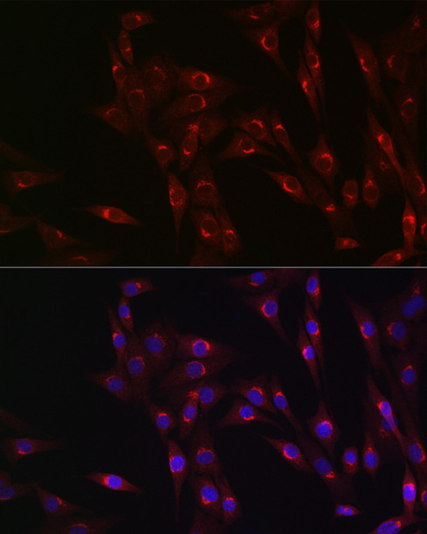
| Host: |
Rabbit |
| Applications: |
WB/IF |
| Reactivity: |
Human/Mouse/Rat |
| Note: |
STRICTLY FOR FURTHER SCIENTIFIC RESEARCH USE ONLY (RUO). MUST NOT TO BE USED IN DIAGNOSTIC OR THERAPEUTIC APPLICATIONS. |
| Short Description: |
Rabbit polyclonal antibody anti-MR1 (23-291) is suitable for use in Western Blot and Immunofluorescence research applications. |
| Clonality: |
Polyclonal |
| Conjugation: |
Unconjugated |
| Isotype: |
IgG |
| Formulation: |
PBS with 0.05% Proclin300, 50% Glycerol, pH7.3. |
| Purification: |
Affinity purification |
| Dilution Range: |
WB 1:1000-1:5000IF/ICC 1:50-1:200 |
| Storage Instruction: |
Store at-20°C for up to 1 year from the date of receipt, and avoid repeat freeze-thaw cycles. |
| Gene Symbol: |
MR1 |
| Gene ID: |
3140 |
| Uniprot ID: |
HMR1_HUMAN |
| Immunogen Region: |
23-291 |
| Immunogen: |
Recombinant fusion protein containing a sequence corresponding to amino acids 23-291 of human MR1 (NP_001522.1). |
| Immunogen Sequence: |
RTHSLRYFRLGVSDPIHGVP EFISVGYVDSHPITTYDSVT RQKEPRAPWMAENLAPDHWE RYTQLLRGWQQMFKVELKRL QRHYNHSGSHTYQRMIGCEL LEDGSTTGFLQYAYDGQDFL IFNKDTLSWLAVDNVAHTIK QAWEANQHELLYQKNWLEEE CIAWLKRFLEYGKDTLQRTE PPLVRVNRKETFPGVTALFC KAHGFYPPEIYMTWMKNGEE IVQEIDYGDILPSGDGTYQ |
| Tissue Specificity | Ubiquitous. Low expression is detected in peripheral blood B cells, T cells, monocytes and in bronchial epithelial cells (at protein level). Expressed in plasmablasts or plasma B cells in the lamina propria of ileum, appendix and colon (at protein level). Highly expressed on a subset of CD45-positive CD3-positive thymocytes (at protein level). |
| Post Translational Modifications | N-glycosylated. |
| Function | Antigen-presenting molecule specialized in displaying microbial pyrimidine-based metabolites to alpha-beta T cell receptors (TCR) on innate-type mucosal-associated invariant T (MAIT) cells. In complex with B2M preferentially presents riboflavin-derived metabolites to semi-invariant TRAV1-2 TCRs on MAIT cells, guiding immune surveillance of the microbial metabolome at mucosal epithelial barriers. Signature pyrimidine-based microbial antigens are generated via non-enzymatic condensation of metabolite intermediates of the riboflavin pathway with by-products arising from other metabolic pathways such as glycolysis. Typical potent antigenic metabolites are 5-(2-oxoethylideneamino)-6-D-ribitylaminouracil (5-OE-RU) and 5-(2-oxopropylideneamino)-6-D-ribitylaminouracil (5-OP-RU), products of condensation of 5-amino-6-D-ribityaminouracil (5-A-RU) with glyoxal or methylglyoxal by-products, respectively. May present microbial antigens to various TRAV1-2-negative MAIT cell subsets, providing for unique recognition of diverse microbes, including pathogens that do not synthesize riboflavin. Upon antigen recognition, elicits rapid innate-type MAIT cell activation to eliminate pathogenic microbes by directly killing infected cells. During T cell development, drives thymic selection and post-thymic terminal differentiation of MAIT cells in a process dependent on commensal microflora. Acts as an immune sensor of cancer cell metabolome. May present a tumor-specific or -associated metabolite essential for cancer cell survival to a pan-cancer TCR consisting of TRAV38.2-DV8*TRAJ31 alpha chain paired with a TRBV25.1*TRBJ2.3 beta chain on a non-MAIT CD8-positive T cell clone (MC.7.G5), triggering T cell-mediated killing of a wide range of cancer cell types. |
| Protein Name | Major Histocompatibility Complex Class I-Related Gene ProteinMhc Class I-Related Gene ProteinClass I Histocompatibility Antigen-Like Protein |
| Cellular Localisation | Cell MembraneSingle-Pass Type I Membrane ProteinEndoplasmic Reticulum MembraneGolgi Apparatus MembraneEarly Endosome MembraneLate Endosome MembraneIn The Absence Of Antigen Remains Within The Endoplasmic Reticulum Where It Acts As A Metabolite SensorAntigen Binding Triggers Trafficking Of The Ternary Complex To The Plasma MembraneAfter PresentationMost Of These Complexes Are Rapidly Internalized And Degraded Via EndocytosisA Small Subset Recycles Via Endosomes Back To The Plasma Membrane And May Thus Acquire And Present New Antigens That Do Not Efficiently Reach The Endoplasmic ReticulumIsoform 1: Cell MembraneSingle-Pass Membrane ProteinIsoform 3: Cell MembraneThe Larger Proportion Remains In The Er In An Immature StateThe Subset That Reach Cell Surface Does It Through A B2m-Independent PathwayIsoform 4: Secreted |
| Alternative Antibody Names | Anti-Major Histocompatibility Complex Class I-Related Gene Protein antibodyAnti-Mhc Class I-Related Gene Protein antibodyAnti-Class I Histocompatibility Antigen-Like Protein antibodyAnti-MR1 antibody |
Information sourced from Uniprot.org
12 months for antibodies. 6 months for ELISA Kits. Please see website T&Cs for further guidance


