| Host: |
Rabbit |
| Applications: |
WB/IHC |
| Reactivity: |
Human/Mouse/Rat |
| Note: |
STRICTLY FOR FURTHER SCIENTIFIC RESEARCH USE ONLY (RUO). MUST NOT TO BE USED IN DIAGNOSTIC OR THERAPEUTIC APPLICATIONS. |
| Short Description: |
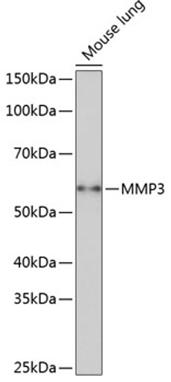
Rabbit monoclonal antibody anti-MMP3 (385-477) is suitable for use in Western Blot and Immunohistochemistry research applications. |
| Clonality: |
Monoclonal |
| Clone ID: |
S7MR |
| Conjugation: |
Unconjugated |
| Isotype: |
IgG |
| Formulation: |
PBS with 0.05% Proclin300, 0.05% BSA, 50% Glycerol, pH7.3. |
| Purification: |
Affinity purification |
| Dilution Range: |
WB 1:500-1:1000IHC-P 1:50-1:200 |
| Storage Instruction: |
Store at-20°C for up to 1 year from the date of receipt, and avoid repeat freeze-thaw cycles. |
| Gene Symbol: |
MMP3 |
| Gene ID: |
4314 |
| Uniprot ID: |
MMP3_HUMAN |
| Immunogen Region: |
385-477 |
| Immunogen: |
Recombinant fusion protein containing a sequence corresponding to amino acids 385-477 of human MMP3 (NP_002413.1). |
| Immunogen Sequence: |
VRKIDAAISDKEKNKTYFFV EDKYWRFDEKRNSMEPGFPK QIAEDFPGIDSKIDAVFEEF GFFYFFTGSSQLEFDPNAKK VTHTLKSNSWLNC |
| Post Translational Modifications | Directly cleaved by HTRA2 to produce active form. |
| Function | Metalloproteinase with a rather broad substrate specificity that can degrade fibronectin, laminin, gelatins of type I, III, IV, and V.collagens III, IV, X, and IX, and cartilage proteoglycans. Activates different molecules including growth factors, plasminogen or other matrix metalloproteinases such as MMP9. Once released into the extracellular matrix (ECM), the inactive pro-enzyme is activated by the plasmin cascade signaling pathway. Acts also intracellularly. For example, in dopaminergic neurons, gets activated by the serine protease HTRA2 upon stress and plays a pivotal role in DA neuronal degeneration by mediating microglial activation and alpha-synuclein/SNCA cleavage. In addition, plays a role in immune response and possesses antiviral activity against various viruses such as vesicular stomatitis virus, influenza A virus (H1N1) and human herpes virus 1. Mechanistically, translocates from the cytoplasm into the cell nucleus upon virus infection to influence NF-kappa-B activities. |
| Protein Name | Stromelysin-1Sl-1Matrix Metalloproteinase-3Mmp-3Transin-1 |
| Database Links | Reactome: R-HSA-1442490Reactome: R-HSA-1474228Reactome: R-HSA-1592389Reactome: R-HSA-2022090Reactome: R-HSA-2179392Reactome: R-HSA-6785807Reactome: R-HSA-9009391 |
| Cellular Localisation | SecretedExtracellular SpaceExtracellular MatrixNucleusCytoplasm |
| Alternative Antibody Names | Anti-Stromelysin-1 antibodyAnti-Sl-1 antibodyAnti-Matrix Metalloproteinase-3 antibodyAnti-Mmp-3 antibodyAnti-Transin-1 antibodyAnti-MMP3 antibodyAnti-STMY1 antibody |
Information sourced from Uniprot.org
12 months for antibodies. 6 months for ELISA Kits. Please see website T&Cs for further guidance


