| Host: |
Chicken |
| Applications: |
WB/IHC/ICC |
| Reactivity: |
Bovine/Human/Mouse/Rat |
| Note: |
STRICTLY FOR FURTHER SCIENTIFIC RESEARCH USE ONLY (RUO). MUST NOT TO BE USED IN DIAGNOSTIC OR THERAPEUTIC APPLICATIONS. |
| Short Description: |
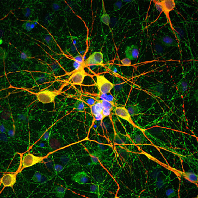
Chicken polyclonal antibody anti-MAP2 is suitable for use in Western Blot, Immunohistochemistry and Immunocytochemistry research applications. |
| Clonality: |
Polyclonal |
| Conjugation: |
Unconjugated |
| Isotype: |
IgY |
| Formulation: |
Total IgY fraction in PBS + 10 mM Sodium Azide. |
| Purification: |
This antibody was total igy fraction. |
| Dilution Range: |
WB 1:20000IHC 1:2500-1:10, 000ICC 1:1000-1:20, 000IP |
| Storage Instruction: |
Store at-20°C for up to 1 year from the date of receipt, and avoid repeat freeze-thaw cycles. |
| Gene Symbol: |
MAP2 |
| Gene ID: |
4133 |
| Uniprot ID: |
MTAP2_HUMAN |
| Immunogen: |
recombinant bovine MAP2 protein expressed in and purification from E. Coli. |
| Post Translational Modifications | Phosphorylated at serine residues in K-X-G-S motifs by MAP/microtubule affinity-regulating kinase (MARK1 or MARK2), causing detachment from microtubules, and their disassembly. Isoform 2 is probably phosphorylated by PKA at Ser-323, Ser-354 and Ser-386 and by FYN at Tyr-67. The interaction with KNDC1 enhances MAP2 threonine phosphorylation. |
| Function | The exact function of MAP2 is unknown but MAPs may stabilize the microtubules against depolymerization. They also seem to have a stiffening effect on microtubules. |
| Protein Name | Microtubule-Associated Protein 2Map-2 |
| Cellular Localisation | CytoplasmCytoskeletonCell ProjectionDendrite |
| Alternative Antibody Names | Anti-Microtubule-Associated Protein 2 antibodyAnti-Map-2 antibodyAnti-MAP2 antibody |
Information sourced from Uniprot.org
12 months for antibodies. 6 months for ELISA Kits. Please see website T&Cs for further guidance


