| Host: |
Rabbit |
| Applications: |
WB/IF |
| Reactivity: |
Human/Mouse/Rat |
| Note: |
STRICTLY FOR FURTHER SCIENTIFIC RESEARCH USE ONLY (RUO). MUST NOT TO BE USED IN DIAGNOSTIC OR THERAPEUTIC APPLICATIONS. |
| Short Description: |
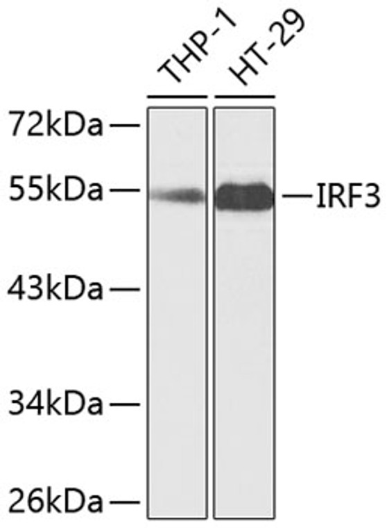
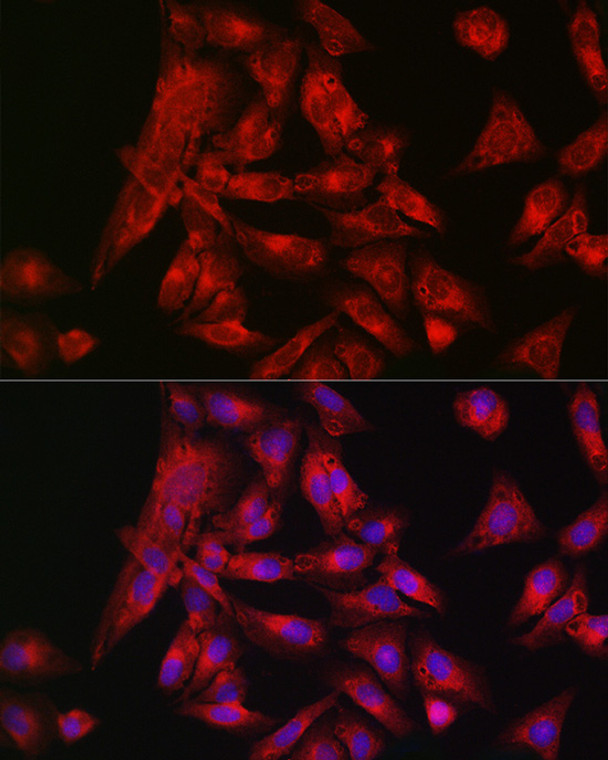
Rabbit polyclonal antibody anti-IRF3 (100-200) is suitable for use in Western Blot and Immunofluorescence research applications. |
| Clonality: |
Polyclonal |
| Conjugation: |
Unconjugated |
| Isotype: |
IgG |
| Formulation: |
PBS with 0.01% Thimerosal, 50% Glycerol, pH7.3. |
| Purification: |
Affinity purification |
| Dilution Range: |
WB 1:500-1:2000IF/ICC 1:50-1:200 |
| Storage Instruction: |
Store at-20°C for up to 1 year from the date of receipt, and avoid repeat freeze-thaw cycles. |
| Gene Symbol: |
IRF3 |
| Gene ID: |
3661 |
| Uniprot ID: |
IRF3_HUMAN |
| Immunogen Region: |
100-200 |
| Immunogen: |
Recombinant fusion protein containing a sequence corresponding to amino acids 100-200 of human IRF3 (NP_001184051.1). |
| Immunogen Sequence: |
PHDPHKIYEFVNSGVGDFSQ PDTSPDTNGGGSTSDTQEDI LDELLGNMVLAPLPDPGPPS LAVAPEPCPQPLRSPSLDNP TPFPNLGPSENPLKRLLVPG E |
| Tissue Specificity | Expressed constitutively in a variety of tissues. |
| Post Translational Modifications | Constitutively phosphorylated on many Ser/Thr residues. Activated following phosphorylation by TBK1 and IKBKE. Innate adapter protein MAVS, STING1 or TICAM1 are first activated by viral RNA, cytosolic DNA, and bacterial lipopolysaccharide (LPS), respectively, leading to activation of the kinases TBK1 and IKBKE. These kinases then phosphorylate the adapter proteins on the pLxIS motif, leading to recruitment of IRF3, thereby licensing IRF3 for phosphorylation by TBK1. Phosphorylated IRF3 dissociates from the adapter proteins, dimerizes, and then enters the nucleus to induce IFNs. Ubiquitinated.ubiquitination involves RBCK1 leading to proteasomal degradation. Polyubiquitinated.ubiquitination involves TRIM21 leading to proteasomal degradation. Ubiquitinated by UBE3C, leading to its degradation. ISGylated by HERC5 resulting in sustained IRF3 activation and in the inhibition of IRF3 ubiquitination by disrupting PIN1 binding. The phosphorylation state of IRF3 does not alter ISGylation. Proteolytically cleaved by apoptotic caspases during apoptosis, leading to its inactivation. Cleavage by CASP3 during virus-induced apoptosis inactivates it, preventing cytokine overproduction. (Microbial infection) ISGylated. ISGylation is cleaved and removed by SARS-COV-2 nsp3 which attenuates type I interferon responses. (Microbial infection) Phosphorylation and subsequent activation of IRF3 is inhibited by vaccinia virus protein E3. (Microbial infection) Phosphorylated by herpes simplex virus 1/HHV-1 US3 at Ser-175 to prevent IRF3 activation. |
| Function | Key transcriptional regulator of type I interferon (IFN)-dependent immune responses which plays a critical role in the innate immune response against DNA and RNA viruses. Regulates the transcription of type I IFN genes (IFN-alpha and IFN-beta) and IFN-stimulated genes (ISG) by binding to an interferon-stimulated response element (ISRE) in their promoters. Acts as a more potent activator of the IFN-beta (IFNB) gene than the IFN-alpha (IFNA) gene and plays a critical role in both the early and late phases of the IFNA/B gene induction. Found in an inactive form in the cytoplasm of uninfected cells and following viral infection, double-stranded RNA (dsRNA), or toll-like receptor (TLR) signaling, is phosphorylated by IKBKE and TBK1 kinases. This induces a conformational change, leading to its dimerization and nuclear localization and association with CREB binding protein (CREBBP) to form dsRNA-activated factor 1 (DRAF1), a complex which activates the transcription of the type I IFN and ISG genes. Can activate distinct gene expression programs in macrophages and can induce significant apoptosis in primary macrophages. In response to Sendai virus infection, is recruited by TOMM70:HSP90AA1 to mitochondrion and forms an apoptosis complex TOMM70:HSP90AA1:IRF3:BAX inducing apoptosis. Key transcription factor regulating the IFN response during SARS-CoV-2 infection. |
| Protein Name | Interferon Regulatory Factor 3Irf-3 |
| Database Links | Reactome: R-HSA-1169408Reactome: R-HSA-1606341Reactome: R-HSA-168928Reactome: R-HSA-3134973Reactome: R-HSA-3134975Reactome: R-HSA-3270619Reactome: R-HSA-877300Reactome: R-HSA-9013973Reactome: R-HSA-909733Reactome: R-HSA-918233Reactome: R-HSA-933541Reactome: R-HSA-936440Reactome: R-HSA-936964Reactome: R-HSA-9692916Reactome: R-HSA-9705671 |
| Cellular Localisation | CytoplasmNucleusMitochondrionShuttles Between Cytoplasmic And Nuclear CompartmentsWith Export Being The Prevailing EffectWhen ActivatedIrf3 Interaction With Crebbp Prevents Its Export To The CytoplasmRecruited To Mitochondria Via Tomm70:Hsp90aa1 Upon Sendai Virus Infection |
| Alternative Antibody Names | Anti-Interferon Regulatory Factor 3 antibodyAnti-Irf-3 antibodyAnti-IRF3 antibody |
Information sourced from Uniprot.org
12 months for antibodies. 6 months for ELISA Kits. Please see website T&Cs for further guidance


