| Host: |
Rabbit |
| Applications: |
WB/IHC/IF |
| Reactivity: |
Human/Mouse/Rat |
| Note: |
STRICTLY FOR FURTHER SCIENTIFIC RESEARCH USE ONLY (RUO). MUST NOT TO BE USED IN DIAGNOSTIC OR THERAPEUTIC APPLICATIONS. |
| Short Description: |
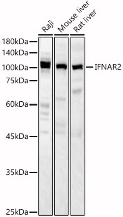
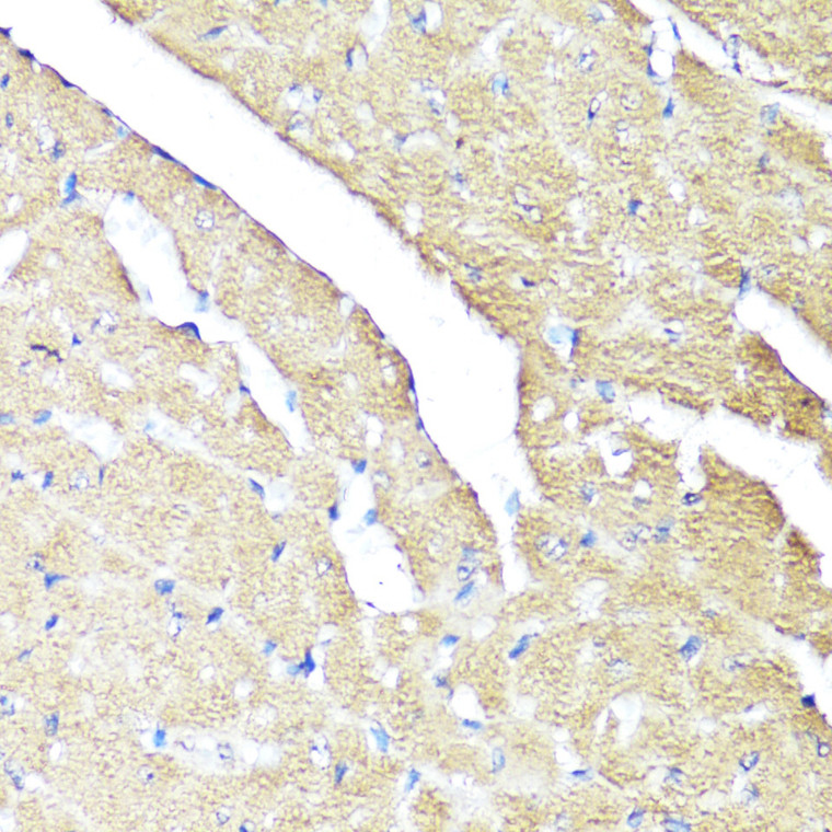
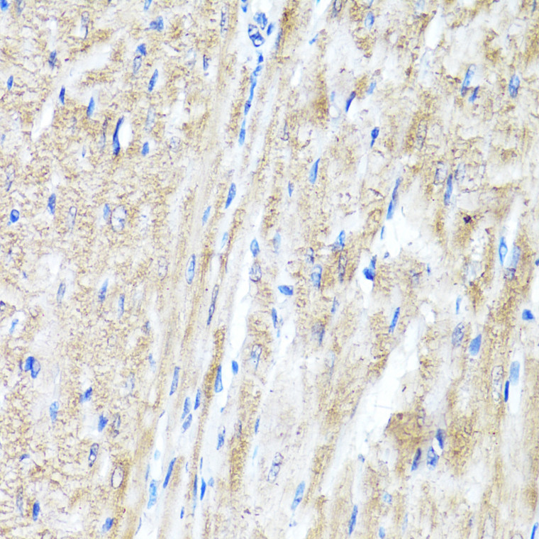
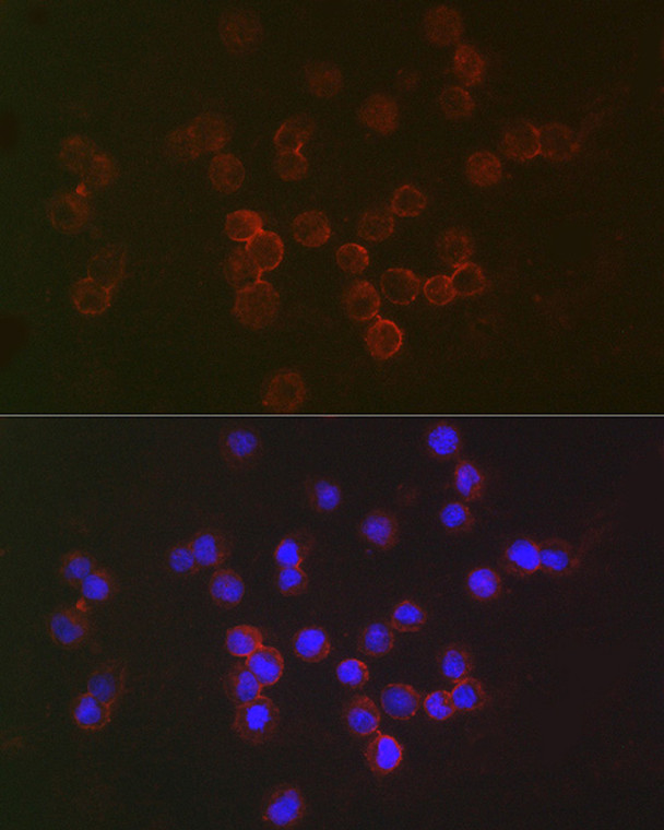
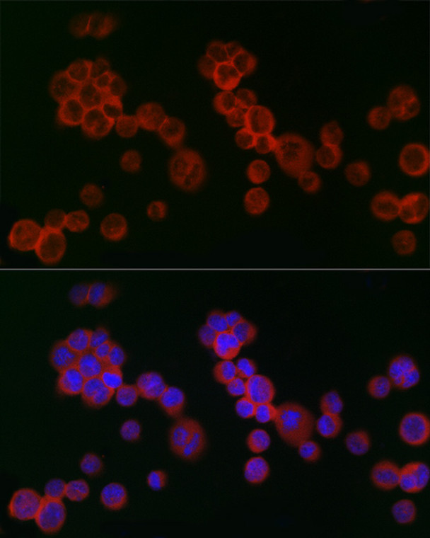
Rabbit polyclonal antibody anti-IFNAR2 (27-515) is suitable for use in Western Blot, Immunohistochemistry and Immunofluorescence research applications. |
| Clonality: |
Polyclonal |
| Conjugation: |
Unconjugated |
| Isotype: |
IgG |
| Formulation: |
PBS with 0.05% Proclin300, 50% Glycerol, pH7.3. |
| Purification: |
Affinity purification |
| Dilution Range: |
WB 1:1000-1:5000IHC-P 1:50-1:100IF/ICC 1:50-1:200 |
| Storage Instruction: |
Store at-20°C for up to 1 year from the date of receipt, and avoid repeat freeze-thaw cycles. |
| Gene Symbol: |
IFNAR2 |
| Gene ID: |
3455 |
| Uniprot ID: |
INAR2_HUMAN |
| Immunogen Region: |
27-515 |
| Immunogen: |
Recombinant fusion protein containing a sequence corresponding to amino acids 27-515 of human IFNAR2 (NP_997468.1). |
| Immunogen Sequence: |
ISYDSPDYTDESCTFKISLR NFRSILSWELKNHSIVPTHY TLLYTIMSKPEDLKVVKNCA NTTRSFCDLTDEWRSTHEAY VTVLEGFSGNTTLFSCSHNF WLAIDMSFEPPEFEIVGFTN HINVMVKFPSIVEEELQFDL SLVIEEQSEGIVKKHKPEIK GNMSGNFTYIIDKLIPNTNY CVSVYLEHSDEQAVIKSPLK CTLLPPGQESESAESAK |
| Tissue Specificity | Isoform 3 is detected in the urine (at protein level). Expressed in blood cells. Expressed in lymphoblastoid and fibrosarcoma cell lines. |
| Post Translational Modifications | Phosphorylated on tyrosine residues upon interferon binding. Phosphorylation at Tyr-337 or Tyr-512 are sufficient to mediate interferon dependent activation of STAT1, STAT2 and STAT3 leading to antiproliferative effects on many different cell types. Glycosylated. |
| Function | Together with IFNAR1, forms the heterodimeric receptor for type I interferons (including interferons alpha, beta, epsilon, omega and kappa). Type I interferon binding activates the JAK-STAT signaling cascade, resulting in transcriptional activation or repression of interferon-regulated genes that encode the effectors of the interferon response. Mechanistically, type I interferon-binding brings the IFNAR1 and IFNAR2 subunits into close proximity with one another, driving their associated Janus kinases (JAKs) (TYK2 bound to IFNAR1 and JAK1 bound to IFNAR2) to cross-phosphorylate one another. The activated kinases phosphorylate specific tyrosine residues on the intracellular domains of IFNAR1 and IFNAR2, forming docking sites for the STAT transcription factors (STAT1, STAT2 and STAT). STAT proteins are then phosphorylated by the JAKs, promoting their translocation into the nucleus to regulate expression of interferon-regulated genes. Isoform 3: Potent inhibitor of type I IFN receptor activity. |
| Protein Name | Interferon Alpha/Beta Receptor 2Ifn-R-2Ifn-Alpha Binding ProteinIfn-Alpha/Beta Receptor 2Interferon Alpha Binding ProteinType I Interferon Receptor 2 |
| Database Links | Reactome: R-HSA-909733 P48551-2Reactome: R-HSA-912694 P48551-2Reactome: R-HSA-9679191 P48551-2Reactome: R-HSA-9705671 P48551-2 |
| Cellular Localisation | Isoform 1: Cell MembraneSingle-Pass Type I Membrane ProteinIsoform 2: Cell MembraneIsoform 3: Secreted |
| Alternative Antibody Names | Anti-Interferon Alpha/Beta Receptor 2 antibodyAnti-Ifn-R-2 antibodyAnti-Ifn-Alpha Binding Protein antibodyAnti-Ifn-Alpha/Beta Receptor 2 antibodyAnti-Interferon Alpha Binding Protein antibodyAnti-Type I Interferon Receptor 2 antibodyAnti-IFNAR2 antibodyAnti-IFNABR antibodyAnti-IFNARB antibody |
Information sourced from Uniprot.org
12 months for antibodies. 6 months for ELISA Kits. Please see website T&Cs for further guidance

