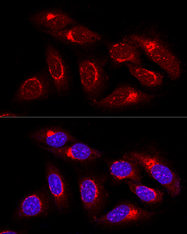
| Host: |
Rabbit |
| Applications: |
WB/IF |
| Reactivity: |
Human |
| Note: |
STRICTLY FOR FURTHER SCIENTIFIC RESEARCH USE ONLY (RUO). MUST NOT TO BE USED IN DIAGNOSTIC OR THERAPEUTIC APPLICATIONS. |
| Short Description: |
Rabbit polyclonal antibody anti-GORASP1 (221-440) is suitable for use in Western Blot and Immunofluorescence research applications. |
| Clonality: |
Polyclonal |
| Conjugation: |
Unconjugated |
| Isotype: |
IgG |
| Formulation: |
PBS with 0.02% Sodium Azide, 50% Glycerol, pH7.3. |
| Purification: |
Affinity purification |
| Dilution Range: |
WB 1:500-1:2000IF/ICC 1:10-1:100 |
| Storage Instruction: |
Store at-20°C for up to 1 year from the date of receipt, and avoid repeat freeze-thaw cycles. |
| Gene Symbol: |
GORASP1 |
| Gene ID: |
64689 |
| Uniprot ID: |
GORS1_HUMAN |
| Immunogen Region: |
221-440 |
| Immunogen: |
Recombinant fusion protein containing a sequence corresponding to amino acids 221-440 of human GRASP65 (NP_114105.1). |
| Immunogen Sequence: |
ALPLGAPPPDALPPGPTPED SPSLETGSRQSDYMEALLQA PGSSMEDPLPGPGSPSHSAP DPDGLPHFMETPLQPPPPVQ RVMDPGFLDVSGISLLDNSN ASVWPSLPSSTELTTTAVST SGPEDICSSSSSHERGGEAT WSGSEFEVSFLDSPGAQAQA DHLPQLTLPDSLTSAASPED GLSAELLEAQAEEEPASTEG LDTGTEAEGLDSQAQISTTE |
| Post Translational Modifications | Phosphorylated by CDC2/B1 and PLK kinases during mitosis. Phosphorylation cycle correlates with the cisternal stacking cycle. Phosphorylation of the homodimer prevents the association of dimers into higher-order oligomers, leading to cisternal unstacking. Target for caspase-3 cleavage during apoptosis. The cleavage contributes to Golgi fragmentation and occurs very early in the execution phase of apoptosis. Myristoylated. |
| Function | Key structural protein of the Golgi apparatus. The membrane cisternae of the Golgi apparatus adhere to each other to form stacks, which are aligned side by side to form the Golgi ribbon. Acting in concert with GORASP2/GRASP55, is required for the formation and maintenance of the Golgi ribbon, and may be dispensable for the formation of stacks. However, other studies suggest that GORASP1 plays an important role in assembly and membrane stacking of the cisternae, and in the reassembly of Golgi stacks after breakdown during mitosis. Caspase-mediated cleavage of GORASP1 is required for fragmentation of the Golgi during apoptosis. Also mediates, via its interaction with GOLGA2/GM130, the docking of transport vesicles with the Golgi membranes. Mediates ER stress-induced unconventional (ER/Golgi-independent) trafficking of core-glycosylated CFTR to cell membrane. |
| Protein Name | Golgi Reassembly-Stacking Protein 1Golgi Peripheral Membrane Protein P65Golgi Phosphoprotein 5Golph5Golgi Reassembly-Stacking Protein Of 65 KdaGrasp65 |
| Database Links | Reactome: R-HSA-162658Reactome: R-HSA-204005Reactome: R-HSA-6807878 |
| Cellular Localisation | Golgi ApparatusCis-Golgi Network MembranePeripheral Membrane ProteinCytoplasmic SideEndoplasmic Reticulum-Golgi Intermediate Compartment Membrane |
| Alternative Antibody Names | Anti-Golgi Reassembly-Stacking Protein 1 antibodyAnti-Golgi Peripheral Membrane Protein P65 antibodyAnti-Golgi Phosphoprotein 5 antibodyAnti-Golph5 antibodyAnti-Golgi Reassembly-Stacking Protein Of 65 Kda antibodyAnti-Grasp65 antibodyAnti-GORASP1 antibodyAnti-GOLPH5 antibodyAnti-GRASP65 antibody |
Information sourced from Uniprot.org
12 months for antibodies. 6 months for ELISA Kits. Please see website T&Cs for further guidance


