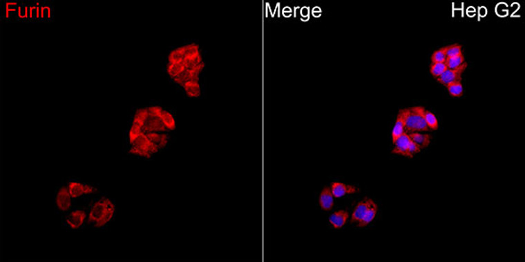
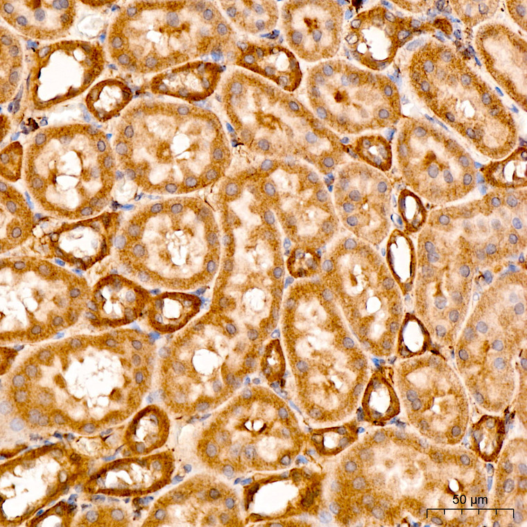
| Host: |
Rabbit |
| Applications: |
WB |
| Reactivity: |
Human/Mouse/Rat |
| Note: |
STRICTLY FOR FURTHER SCIENTIFIC RESEARCH USE ONLY (RUO). MUST NOT TO BE USED IN DIAGNOSTIC OR THERAPEUTIC APPLICATIONS. |
| Short Description: |
Rabbit polyclonal antibody anti-FURIN (700-794) is suitable for use in Western Blot research applications. |
| Clonality: |
Polyclonal |
| Conjugation: |
Unconjugated |
| Isotype: |
IgG |
| Formulation: |
PBS with 0.02% Sodium Azide, 50% Glycerol, pH7.3. |
| Purification: |
Affinity purification |
| Dilution Range: |
WB 1:500-1:2000 |
| Storage Instruction: |
Store at-20°C for up to 1 year from the date of receipt, and avoid repeat freeze-thaw cycles. |
| Gene Symbol: |
FURIN |
| Gene ID: |
5045 |
| Uniprot ID: |
FURIN_HUMAN |
| Immunogen Region: |
700-794 |
| Immunogen: |
A synthetic peptide corresponding to a sequence within amino acids 700-794 of human Furin (NP_002560.1). |
| Immunogen Sequence: |
AGQRLRAGLLPSHLPEVVAG LSCAFIVLVFVTVFLVLQLR SGFSFRGVKVYTMDRGLISY KGLPPEAWQEECPSDSEEDE GRGERTAFIKDQSAL |
| Tissue Specificity | Seems to be expressed ubiquitously. |
| Post Translational Modifications | The inhibition peptide, which plays the role of an intramolecular chaperone, is autocatalytically removed in the endoplasmic reticulum (ER) and remains non-covalently bound to furin as a potent autoinhibitor. Following transport to the trans Golgi, a second cleavage within the inhibition propeptide results in propeptide dissociation and furin activation. Phosphorylation is required for TGN localization of the endoprotease. In vivo, exists as di-, mono- and non-phosphorylated forms. |
| Function | Ubiquitous endoprotease within constitutive secretory pathways capable of cleavage at the RX(K/R)R consensus motif. Mediates processing of TGFB1, an essential step in TGF-beta-1 activation. Converts through proteolytic cleavage the non-functional Brain natriuretic factor prohormone into its active hormone BNP(1-32). By mediating processing of accessory subunit ATP6AP1/Ac45 of the V-ATPase, regulates the acidification of dense-core secretory granules in islets of Langerhans cells. (Microbial infection) Cleaves and activates diphtheria toxin DT. (Microbial infection) Cleaves and activates anthrax toxin protective antigen (PA). (Microbial infection) Cleaves and activates HIV-1 virus Envelope glycoprotein gp160. (Microbial infection) Required for H7N1 and H5N1 influenza virus infection probably by cleaving hemagglutinin. (Microbial infection) Able to cleave S.pneumoniae serine-rich repeat protein PsrP. (Microbial infection) Facilitates human coronaviruses EMC and SARS-CoV-2 infections by proteolytically cleaving the spike protein at the monobasic S1/S2 cleavage site. This cleavage is essential for spike protein-mediated cell-cell fusion and entry into human lung cells. (Microbial infection) Facilitates mumps virus infection by proteolytically cleaving the viral fusion protein F. |
| Protein Name | FurinDibasic-Processing EnzymePaired Basic Amino Acid Residue-Cleaving EnzymePace |
| Database Links | Reactome: R-HSA-1181150Reactome: R-HSA-1442490Reactome: R-HSA-1566948Reactome: R-HSA-1592389Reactome: R-HSA-159782Reactome: R-HSA-167060Reactome: R-HSA-171286Reactome: R-HSA-186797Reactome: R-HSA-1912420Reactome: R-HSA-2173789Reactome: R-HSA-2173796Reactome: R-HSA-5210891Reactome: R-HSA-6809371Reactome: R-HSA-8963889Reactome: R-HSA-9662834Reactome: R-HSA-9679191Reactome: R-HSA-9694614Reactome: R-HSA-9733458Reactome: R-HSA-977225 |
| Cellular Localisation | Golgi ApparatusTrans-Golgi Network MembraneSingle-Pass Type I Membrane ProteinCell MembraneSecretedEndosome MembraneShuttles Between The Trans-Golgi Network And The Cell SurfacePropeptide Cleavage Is A Prerequisite For Exit Of Furin Molecules Out Of The Endoplasmic Reticulum (Er)A Second Cleavage Within The Propeptide Occurs In The Trans Golgi Network (Tgn)Followed By The Release Of The Propeptide And The Activation Of Furin |
| Alternative Antibody Names | Anti-Furin antibodyAnti-Dibasic-Processing Enzyme antibodyAnti-Paired Basic Amino Acid Residue-Cleaving Enzyme antibodyAnti-Pace antibodyAnti-FURIN antibodyAnti-FUR antibodyAnti-PACE antibodyAnti-PCSK3 antibody |
Information sourced from Uniprot.org
12 months for antibodies. 6 months for ELISA Kits. Please see website T&Cs for further guidance


